И нет, свидетельств того, что ортодонтическое "лечение" способствует здоровью дёсен/пародонта — нет.
Содержание: https://healthy-back.livejournal.com/436663.html#Cont (https://healthy-back.dreamwidth.org/424733.html#Cont)
Назад: https://healthy-back.livejournal.com/455638.html (https://healthy-back.dreamwidth.org/442656.html)
Вперёд: Протокол именно для коррекции нарушений сна Stanford Protocol / Стэнфордский (Стенфордский) протокол https://healthy-back.livejournal.com/462192.html (https://healthy-back.dreamwidth.org/448267.html)
UPD 12/08/2023: См.
kaoruniten Мой (вероятно) неудачный опыт лечения брекетами
https://pikabu.ru/story/moy_veroyatno_neudachnyiy_opyit_lecheniya_breketami_10524314?utm_source=linkshare&utm_medium=sharing

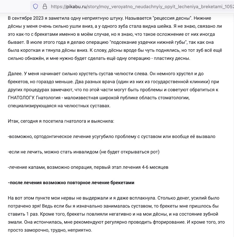
И первый коммент от llapa.lapeonia к этой истории
https://pikabu.ru/story/moy_veroyatno_neudachnyiy_opyit_lecheniya_breketami_10524314#comments

1) https://ronald-ead.squarespace.com/new-blog-1/2018/1/31/the-best-adult-palate-expander (https://healthy-back.livejournal.com/387453.html, https://healthy-back.dreamwidth.org/379343.html)
2) Конспект диссертации Махортовой - I https://healthy-back.livejournal.com/459796.html (https://healthy-back.dreamwidth.org/446353.html)
побочные эффекты расширения нёба
— вестибулярный наклон опорных зубов,
— фенестрации вестибулярной кортикальной пластинки,
— резорбцию корней и
— рецессии десны [7, 10, 17, 24, 32, 33, 58, 81, 86-89, 196]
7. Арсенина О.И. Комплексная диагностика и лечение дистальной окклюзии зубных рядов несъёмной ортодонтической техникой / Арсенина О. И., Надточий А. Г., Попова А. В. -616.314-089.23. -Москва. - 2009. - 70-96.
10. Арсенина О. И. Комплексная диагностика и лечение пациентов с сужением и деформацией верхней челюсти / О. И. Арсенина, Н. В. Попова, П. И. Махортова, Л. А. Гайрбекова // Клиническая стоматология. -2019. №1(89). - 51-57.
17. Гусарина Е.И. Оценка эффективности ортодонтического лечения с помощью обследования на конусно-лучевом компьютерном томографе у пациентов с зубочелюстными аномалиями и особенностями строения тканей пародонта (Часть I) / Гусарина Е.И., Зубарева А.А., Чибисов М.А. // Институт Стоматологии. - Спб., 2016.-С.58-62.
24. Дробышев A.Ю., Дробышева H.С., Клипа И.А., Глушко A.В., Водахова A. Алгоритм диагностики и лечения пациентов с аномалиями челюстей, сопровождающихся сужением верхней челюсти // XXth Congress of the European Association for Cranio-Maxillo-Facial Surgery. Abstract book. Bruges (Belgium), September 14 – 18th. – 2010. – P. 781.
32. Кожевникова Л.А. Сравнительный анализ методик расширения верхней челюсти с использованием ортодонтических аппаратов / Кожевникова Л.А., Джабраилова Г.Д., Абдулкеримова С.М., Керимова К.Н., Пильщикова О.В., Слонова В.М., Геворкян А.А. // Эндодонтия Today. 2019. Т. 17. №4. С. 58-62.
33. Комелягин Д. Ю. Устранение сужения челюстей методом компрессионно-дистракционного остеосинтеза / Д. Ю. Комелягин, Ан.В. Дергаченко, О.З. Топольницкий, А.Б. Слабковская, С.А. Дубин, Ф.И. Владимиров, А.В. Петухов, А.В. Дергаченко, Е.В. Стрига, В.Г. Слипенко, Л.А. Крашенинников, С.В. Яматина, А.В. Пасечников, Х.Я. Вафина, И.А. Строгонов // Голова и шея. 2017;1:37-46.
58. Польма Л.В. Быстрое расширение верхней челюсти в комплексном лечении взрослых пациентов с трансверсальными аномалиями окклюзии/ Польма Л.В., Дробышев А.Ю., Дубова Л.В., Оборотистов Н.Ю. // Ортодонтия. 2006. №3. С. 36-41.
81. Токаревич И.В. Оценка параметров скелетного расширения верхней челюсти аппаратами с разными типом опоры / Токаревич И.В., Хомич А.С. // Современная стоматология. 2018. №2(71). С. 44-50.
86. Фурцев Т. В. Сравнительное исследование поверхностей трех типов имплантатов (TiUnite, SLA, RBM) с контрольным образцом, периимплантитом, обработанных лазером Er;Cr;YSGG длиной волны 2780 нм / Т. В. Фурцев, Г. М. Зеер // Стоматология. 2019;98(3): 52-55
87. Хватова В.А. Функциональная окклюзия в норме и патологии. – М., 1993.-160 с.
88. Хватова В.А. Диагностика и лечение нарушений функциональной окклюзии: Руководство.- Н.Новгород.-1996. – 276с.
89. Хорошилкина Ф.Я., Малыгин Ю.М., Биллиг В.А., Берсенева Е.Л. Сравнительный телерентгенологический анализ строения лицевого скелета в период сменного и постоянного прикуса в норме // Вопросы стоматологии. ЦОЛИУВ. 1979. - С.121-125.
196. Ong SC, Khambay BS, McDonald JP, Cross DL, Brocklebank LM, Ju X. The novel use of three-dimensional surface models to quantify and visualise the immediate changes of the mid-facial skeleton following rapid maxillary expansion. Surgeon 2015;13:132-8.
комбинированное ортодонто-хирургическое лечение с хирургически ассистированным быстрым небным расширением: ряд осложнений, которые могут возникнуть в послеоперационном периоде:
— выраженное кровотечение,
— рецессия десны,
— резорбция корней,
— повреждение ветвей верхнечелюстного нерва,
— послехирургическая боль,
— гибель пульпы зубов,
— периодонтальные проблемы,
— экструзия опорных зубов,
— воспаление кортикальной пластинки,
— синуситы,
— асимметричное расширение,
— отклонение носовой перегородки и
— рецидив деформации [106, 107, 117-119].
106. Baysal A., Karadede I., Hekimoglu S. Evaluation of root resorption following rapid maxillary expansion using cone-beam computed tomography // Angle Orthod. 2012. V.82. P.488-494
107. Baysal A., Uysal T., Veli I., Ozer T. Evaluation of alveolar bone loss following rapid maxillary expansion using cone-beam computed tomography // Korean J Orthod. 2013. V.43. P.83-95.
117. Brunetto DP, Sant’Anna EF, Machado AW, Moon W. Non-surgical treatment of transverse deficiency in adults using Microimplant assisted Rapid Palatal Expansion (MARPE). Dent Press J Orthod 2017;22:110-25.
118. Byloff FK, Mossaz CF. Skeletal and dental changes following sur- gically assisted rapid palatal expansion. Eur J Orthod 2004;26: 403-9.
119. Cameron C.G. Long-Term Effects Of Rapid Maxillary Expansion: A Postero-Anterior Cephalometric Evaluation / Cameron C.G., Franchi L., Bacetti T., Mcnamara J.A. Et Al. // Am. J. Orthod. Dentofacial. Orthop.-2002.-Vol. 121.P.129-135.
4) https://www.sciencedirect.com/science/article/abs/pii/S1761722711000945 (https://pubmed.ncbi.nlm.nih.gov/22257702/)
Gingival recession and adult orthodontics: A clinical evidence-based treatment proposal https://doi.org/10.1016/j.ortho.2011.09.013
The presence of a gingival recession prior to orthodontic treatment is a real problem. Patients are concerned about losing their teeth but may also complain of their unpleasant appearance or root sensitivity in the exposed area. The orthodontist is not sure whether orthodontic treatment can be performed or whether the tooth movement will not aggravate the recession and whether periodontal surgery needs to be done before or after orthodontic treatment. The aim of this paper is to present recent data from the literature and several clinical situations in adults in order to submit a treatment sequence and clarify the role of different periodontal plastic surgery root coverage procedures.
Gingival recession – or, more accurately, periodontal recession since the four components of the periodontium migrate apically – is a frequently observed clinical situation which, in a West European population, affects 60% of young people under 20 years and 90% of adults over 50 (Loë et al.) [1].
The etiology of gingival recession is often multifactorial and, according to Rodier [2], 17% of recessions have no obvious clinical etiology.
5) https://pubmed.ncbi.nlm.nih.gov/22908075/ Eur J Esthet Dent. 2012 Autumn;7(3):266-80.
Combined orthodontic - mucogingival treatment of a deep post-orthodontic gingival recession
Giovanni Zucchelli 1 , Serena Incerti Parenti, Gino Ghigi, Giulio Alessandri Bonetti
Abstract
In this article, the interdisciplinary management of an isolated-type recession defect in a severely compromised mandibular incisor of a young post-orthodontic patient is described. The prognosis of root coverage surgery was very questionable and unpredictable due to the severe root malposition (III Miller class gingival recession).
6) https://link.springer.com/article/10.1007/s00056-018-0159-8
Prevalence of gingival recession after orthodontic treatment of infraversion and open bite
Prävalenz von Gingivarezessionen nach kieferorthopädischer Behandlung von Infraposition und offenem Biss
Juan-Juan Ji, Xu-Dong Li, Qun Fan, Xiao-Jun Liu, Shuang Yao, Zhi Zhou, Shuang Yang & Yong Shen
Journal of Orofacial Orthopedics / Fortschritte der Kieferorthopädie volume 80, pages 1–8 (2019) Cite this article
Results
The prevalence of gingival recession in patients with infraversion and open bite after orthodontic treatment were 80.6 and 75.0%, respectively; these values were 43.4 and 47.5% before treatment, respectively. Notably, the Miller index of gingival recession increased after orthodontic treatment (P < 0.05). The risk of gingival recession in patients with infraversion or open bite after orthodontic treatment was remarkably higher than the risk in other patients (odds ratio [OR] = 16.712 and 5.073, respectively); the gingival recession rate was related to treatment with tooth extraction (OR = 2.043), as well as gingival biotype (OR = 0.341) and gingival index (GI) before orthodontic treatment (OR = 97.404; P < 0.05).
Conclusions
Patients with these two types of low occlusal function are more likely to exhibit gingival recession after orthodontic treatment. Moreover, the prevalence of gingival recession after orthodontic treatment is higher among patients who have undergone tooth extraction during orthodontic treatment, and among those who exhibit thin gingival biotype and high gingival index before orthodontic treatment.
7) https://www.ncbi.nlm.nih.gov/pmc/articles/PMC4149153/ J Clin Diagn Res. 2014 Jul; 8(7): ZD05–ZD07. Published online 2014 Jul 20. doi: 10.7860/JCDR/2014/9767.4555 PMCID: PMC4149153 PMID: 25177647
Management of Gingival Recession Associated with Orthodontic Treatment: A Case Report
Tarun Kumar Rana,1 Megha Phogat, corresponding author Tarun Sharma, Narayana Prasad, and Shailendra Singh
Many patients undergo orthodontic treatment for aesthetic improvement. It is well established that the patients who undergo orthodontic treatment have a high susceptibility to present plaque accumulation on their teeth because of the presence of brackets, wires and/or other orthodontic elements on the teeth surfaces with which the oral hygiene procedures might be more difficult. The orthodontic treatment is a double-action procedure regarding the periodontal tissues which may be very meaningful in increasing the periodontal health status and may be a harmful procedure which can be followed by several types of periodontal complications. There is a strong correlation between the severity and extent of gingival recessions and the orthodontic treatment suggesting that orthodontic tooth movement may lead to gingival recession. The principal objective in the treatment of gingival recession is to cover the exposed root surfaces to improve aesthetics and to reduce hypersensitivity. Different soft tissue grafting procedures have been proposed in the treatment of gingival recessions. Subepithelial connective tissue graft is a reliable method for treatment of gingival recession. The purpose of this case report was to illustrate the relationship between orthodontic therapy and gingival recession and to describe the management of this case.
8) https://meridian.allenpress.com/angle-orthodontist/article/83/6/1093/132322/Gingival-recession-can-orthodontics-be-a-cure
Gingival recession—can orthodontics be a cure? Evidence from a case presentation
William M. Northway Angle Orthod (2013) 83 (6): 1093–1101. https://doi.org/10.2319/012413-76.1
In 2008, the featured cover article in the Journal of the American Dental Association was an exhaustive review of the literature that found that orthodontics provided little benefit to the periodontium; in fact, the net response was small but detrimental.
The objective of this article is to discuss the relationship between the development of a healthy periodontium and the potential impact of orthodontics, to advocate for modifications in orthodontic treatment that might improve the periodontal prognosis, and to present a treatment report that demonstrates concomitant orthodontic and periodontal benefits.
More recently, there has been discussion of a genetically based susceptibility that is manifest in various types of gingival integrity, known as biotype (thick or thin). 10
10. Pontoriero R, Carnevale G. Surgical crown lengthening: a 12-month clinical wound healing study. J Periodontol. 2001;72:841–848.
In 1977, Bernimoulin and Curilovic [11] debunked the correlation between GR and tooth mobility and the correlation between tooth mobility and alveolar bone dehiscence. While they found a positive, significant correlation between GR and bone dehiscence, they questioned the impact of trauma from occlusion.
11. Bernimoulin J, Curilovic Z. Gingival recession and tooth mobility. J Clin Periodontol. 1977; 4:107–114. https://pubmed.ncbi.nlm.nih.gov/266503/
More germane to the realm of orthodontics, recession can occur during development in the form of insufficient space for the dentition on an alveolar ridge. As the discrepancy between tooth mass and available space increases, teeth become more crowded and are ultimately pushed off the ridge, compromising the supporting bone. Thus, the likelihood of recession increases. The process begins in the form of dehiscences or fenestrations. Once these have formed, the absence of alveolar bone makes it more difficult for healthy gingival tissue to persist in the form of a protective barrier.
The hypothesis he proposed was that orthodontic movement in the direction of risk would likely exacerbate the periodontal status and cause GR. In his study, he found that more than 75% of the patients who have received premolar extraction “demonstrate clinically significant gingival recession, leading him to conclude that these patients were compromised from the outset by reduced RSBI."
Daprile et al.9 reported on a 5-year study of dental students, wherein the number of subjects with at least one site of recession increased significantly—as did the total number of foci. What's more, the percentage of affected sites increased with the level of oral hygiene education, and this increase was despite a reduction in harmful dental hygiene habits.
Of the 174 fifteen-year-olds in the sample studied by Bjorn et al.,14 an astounding 62% showed some degree of GR on the labial of maxillary teeth. Interestingly, the group with an “unspecific” toothbrushing technique had fewer lesions than the group that used the “roll or vibratory technique.” The authors speculated that the difference might not have been the technique but the fact that the unspecific group was paying less attention to their oral hygiene.
Loe et al.15 compared samples from Norway and Sri Lanka and found that more than 60% of Norwegian children had recession by age 20 (compared with 30% in Sri Lanka), and that increased to more than 90% at age 50 (compared with 100% of Sri Lankans by age 40). As the Norwegian recession was largely on the buccal and that of the Sri Lankan cohort was more generally distributed, the researchers conjectured that several factors were involved, that much of the Norwegian recession seemed to be more related to mechanical abrasion associated with excessive hygiene systems as opposed to recession caused more by periodontal disease in the Sri Lankan sample.
9) https://www.aegisdentalnetwork.com/cced/2022/03/the-impact-of-orthodontic-retainers-on-gingival-recession-a-best-evidence-review
Compendium March 2022 Volume 43, Issue 3 Peer-Reviewed
The Impact of Orthodontic Retainers on Gingival Recession: A Best-Evidence Review
Nada M. Souccar, DDS, MS; Rawan Oueis, DDS; John Paul Mussleman, Jr., MLIS; Nicolaas C. Geurs, DDS, MS; and Ramzi V. Abou-Arraj, DDS, MS
This best-evidence article reviews the effects of the different types of orthodontic retention appliances, fixed and removable, on the development and progression of gingival recession at the mandibular anterior teeth. Searched databases included PubMed, Scopus, Cochrane Library, Embase, and Dentistry & Oral Sciences. Eleven qualifying publications, including retrospective, prospective, and cross-sectional studies, were included in this review. These studies either did not demonstrate an association between orthodontic retainers and gingival recession or reported that the resulting recession defects were minimal when an association was shown.
An important consideration is that recession could be a late finding following the placement of a retainer and, therefore, may be incipient or absent in short-term evaluations. Prospective studies that specifically address the role that properly positioned fixed retainers may have on gingival recession are needed before a definitive conclusion can be generalized with regard to recommended retention protocols. Factors such as duration of retainer use, number of bonded teeth, and position of fixed retainers relative to their proximity to gingival tissues are not fully elucidated but may have influencing roles on gingival recession. The use of retainers should be based on orthodontic indications to maintain a stable dental arch form, esthetics, and occlusion. Effective oral hygiene and follow-up regimens remain the gold standard in maintaining periodontal health and preventing gingival recession.
Long-term post-orthodontic follow-up, spanning 10 to 20 years, has revealed that less than 30% of cases showed stability in the anterior mandibular segment with 20% of those cases showing likelihood for crowding years after retainer removal.
Gingival recession results in apical migration of the gingival tissue that leads to exposure of the root apical to the cementoenamel junction.18
Recession increases with age and equally affects individuals with high and low levels of oral hygiene.19
Recession can occur labially or lingually, is unesthetic, and can potentially lead to dentin hypersensitivity, root caries, soft-tissue discomfort, and a greater susceptibility to inflammatory insult.20 These clinically adverse conditions highlight the importance of having a proper understanding of the effects that different types of retainers can have on the prevalence of recession in the gingival tissue.
Predisposing factors that have been implicated in the development or progression of gingival recession include
— periodontitis,
— periodontal therapy,
— thin periodontal phenotype,21,22
— improper toothbrushing through increased duration, frequency, force, or brush bristle size,23,24
— presence of intracrevicular restorative margins especially in the absence of keratinized gingiva,21
— orthodontic treatment,25,26 and
— shallow vestibular depth and
— frenum position that interfere with proper plaque control.27
Several of these factors are considered to have a low level of evidence.28
25. Bollen AM, Cunha-Cruz J, Bakko DW, et al. The effects of orthodontic therapy on periodontal health: a systematic review of controlled evidence. J Am Dent Assoc. 2008;139(4):413-422.
26. Joss-Vassalli I, Grebenstein C, Topouzelis N, et al. Orthodontic therapy and gingival recession: a systematic review. Orthod Craniofac Res. 2010;13(3):127-141.
Fixed retainers, which are commonly used to alleviate the need for patient compliance, have been found to have no significant effect on the prevalence of recession regardless of type of wire and bonding technique.7,12,15 Furthermore, the length of retainer use can also be a confounding factor. There is no evidence that has determined an ideal retention duration; however, there is general consensus that retention is recommended for life. One study comparing the prevalence of recession in patients with long-term and short-term use of fixed retainers noted an increase in gingival recession in the long-term group.9 However, these recessions were found mostly buccally, thus weakening the association of the lingually placed fixed retainer on the occurrence of gingival recession.
Despite the general consensus, a study that compared patients with fixed retainers to patients who did not undergo orthodontic treatment had contradictory results stating that the fixed retainer significantly impacted the likelihood of recession by increasing that risk by a 4.8-fold.10 The recessions in that study were labial, and the authors noted that it was unclear whether the recessions occurred as a result of orthodontic treatment or presence of the fixed retainer.
Clinical Conclusion
Within the limitations of the reviewed studies, it is unclear whether fixed or removable orthodontic retainers affect the initiation or progression of marginal gingival recession. Factors such as duration of retainer use, number of bonded teeth, and position of fixed retainers relative to their proximity to gingival tissues are not fully elucidated but may have influencing roles on gingival recession. The use of retainers is dependent on the patient's malocclusion and should be based on orthodontic indications to maintain a stable dental arch form, esthetics, and occlusion.
10) https://pubmed.ncbi.nlm.nih.gov/27868202/ Aust Dent J. 2017 Mar;62 Suppl 1:86-96. doi: 10.1111/adj.12486.
Potential risks of orthodontic therapy: a critical review and conceptual framework M Wishney 1-2 Affiliations PMID: 27868202
This review examines some of the potential risks of orthodontic therapy along with their evidence base. The risks of orthodontic treatment include
— periodontal damage,
— pain,
— root resorption,
— tooth devitalization,
— temporomandibular disorder,
— caries,
— speech problems and
— enamel damage.
These risks can be understood to arise from a synergy between treatment and patient factors. In general terms, treatment factors that can influence risk include appliance type, force vectors and duration of treatment whilst relevant patient factors are both biological and behavioural. Hence, the natural variation between orthodontic treatment plans and patients gives rise to variations in risk. A good understanding of these risks is required for clinicians to obtain informed consent before starting treatment as well as to reduce the potential for harm during treatment. After considering each of these risks, a conceptual framework is presented to help clinicians better understand how orthodontic risks arise and may therefore be mitigated.
11) https://www.ncbi.nlm.nih.gov/pmc/articles/PMC4998922/
Medicine (Baltimore). 2016 Mar; 95(10): e3080. Published online 2016 Mar 11. doi: 10.1097/MD.0000000000003080
Does Orthodontic Treatment Affect the Alveolar Bone Density?
Jian-Hong Yu, DDS, PhD, Heng-Li Huang, PhD, Chien-Feng Liu, DDS, Jay Wu, PhD, Yu-Fen Li, PhD, Ming-Tzu Tsai, PhD, and Jui-Ting Hsu, PhD
During orthodontic tooth movement, the alveolar bone density around the teeth was reduced. However, after a period of bone recovery, the reduced bone density recovered to its previous state from before the orthodontic treatment. However, the bone density around ∼10% of the teeth in this region could not recover to 80% of its state from before the orthodontic treatment.
12) https://www.ncbi.nlm.nih.gov/pmc/articles/PMC4072383/ J Orthod Sci. 2013 Jul-Sep; 2(3): 73–86. doi: 10.4103/2278-0203.119678 PMCID: PMC4072383 PMID: 24987646
Iatrogenic possibilities of orthodontic treatment and modalities of prevention
Nazeer Ahmed Meeran
Orthodontic treatment can play an important role in enhancing esthetics, function, and self-esteem in patients. However, it carries with it the risks of enamel demineralization, tissue damage, root resorption, open gingival embrasures in the form of triangular spaces, allergic reactions to nickel, and treatment failure in the form of relapse. These potential complications are easily avoidable by undertaking certain precautions and timely interventions by both the orthodontist and the patient. The orthodontist must ensure that the patient is aware of the associated risks and stress the importance of the patient's role in preventing these untoward outcomes. The decision whether to proceed with the orthodontic treatment is essentially a risk-benefit analysis, where the perceived benefits of commencing treatment outweigh the potential risks. This article provides an overview of the iatrogenic possibilities of orthodontic treatment and the role of the patient as well as the orthodontist in preventing the associated risks.
Decalcification
Shannon[3] recognized orthodontic patients to be at a higher risk of decalcification or caries. An orthodontic appliance could not, within itself, be a cause of caries. However, oral hygiene problems do occur when fixed appliances are worn. Meticulous attention to oral hygiene is mandatory during the entire treatment period to avoid the risk of enamel decalcification. Banded or bonded teeth, exhibited significantly more white spot lesions compared to the controls without braces.[4] Ogaard[5] noticed that even 5 years after completing the treatment, orthodontic patients had a significantly higher incidence of enamel opacities than untreated controls.
ENAMEL FRACTURES DURING DEBONDING
Improper debonding of orthodontic brackets, particularly ceramic brackets, can result in enamel surface cracks.[25] They can provide stagnation areas for the development of caries, cause partial tooth fracture, or may cause unaesthetic discoloration. Zachrisson[25] found higher prevalence of cracks in debonded teeth compared to untreated teeth. There were appreciably more cracks with chemically bonded ceramic brackets.[26]
POTENTIAL ADVERSE EFFECTS TO THE PERIODONTAL TISSUES
Gingival Inflammation
In many orthodontic patients, the principal reason for the associated gingival and periodontal inflammation involves mechanical irritation caused by the band or cement, in addition to trapped plaque.[36,37] The risk of attachment loss can be anticipated when such iatrogenic irritations are present.[38]
preexisting untreated periodontal disorders, fixed orthodontic appliances and tooth movement can contribute to significant and permanent periodontal damage.[40]
Gingival Recession
Gingival recession has been known to occur as an adverse effect during the orthodontic treatment or after treatment completion and has been noted more frequently during buccal orthodontic tooth movements.[62] If teeth having thin tissue are going to be moved lingually, there is a potential for the tissue to move coronally and become thicker.[63] It is generally advisable to monitor areas of thin gingival tissues periodically as the width of the attached gingiva generally increases with normal growth from the mixed to the permanent dentition.[64]
It has been found that most cases of gingival recession which occur during an orthodontic treatment occurred in the regions of the upper and lower anterior teeth.[65,66,67] The relationship between orthodontic movements and gingival recession has been controversial in relation to tipping movements. Batenhorst et al.[68] found an association between gingival recessions and orthodontic tipping tooth movements of the lower incisors in monkeys. However, other studies revealed no association between gingival recession or mucogingival defects after orthodontic tipping of the incisors.[69,70]
Black Triangles
Black triangle or open gingival embrasure can occur as potential complication in about than 1/3 of all adult orthodontic patients and should be discussed with patients prior to initiating orthodontic treatment.[73,75]
ROOT RESORPTION
A genetic predisposition to root resorption has been recognized recently.[83] Decreased IL-1 production in the case of IL-1B allele 1 may result in relatively less catabolic bone modeling (resorption) at the cortical bone interface with the PDL, which may result in prolonged stress concentrated in the root of the tooth, triggering a cascade of fatigue-related events subsequently leading to root resorption.[83]
PULP DAMAGE AND LOSS OF TOOTH VITALITY
Various studies of changes in pulp tissue vascularity during orthodontic treatment, suggest that blood flow to the dental pulp decreases initially after orthodontic force application. However, it increases thereafter until it reaches a peak 7 days after the application of orthodontic force.[93,94,95,96] These processes, depending on the degree of their disturbances, may cause changes in the metabolic cell activity, cell damage, or defense reactions. Orthodontic forces affect the dental pulp inducing vascular changes that are inflammatory in nature.[97,98] As demonstrated in the rat model, the inflammatory vascular reactions subside within 3 weeks in all tissues.[93,96]
Orthodontic patients may suffer from transient pulp ischemia, causing pain, and discomfort in the first few days after activation of an appliance. This usually settles within a week although pulp death following orthodontic treatment is occasionally reported.[99]
DENTIN HYPERSENSITIVITY AFTER INTERPROXIMAL ENAMEL REDUCTION
DAMAGE TO THE INTRA-ORAL TISSUES
Direct Damage by Removable or Fixed Components
Soft Tissue Complications Related to Micro Implants
ENAMEL ABRASION
HEADGEAR INJURIES
ACCIDENTAL SWALLOWING OF APPLIANCE COMPONENTS
ALLERGIC REACTIONS FROM ORTHODONTIC APPLIANCES
PROLONGATION IN THE TREATMENT DURATION
TREATMENT FAILURE AND RELAPSE
Failure to complete a course of orthodontic treatment is frustratingly common[159] (4-23%). This may either be due to the patient's insistence on removing the appliance earlier for personal reasons like marriage or the orthodontists’ opinion that further continuation of treatment may jeopardize the health of the dentition and the periodontal ligament, in the face of severe root resorption. Sometimes the patient might not be able to maintain oral hygiene in a satisfactory way as expected, resulting in worsening of the periodontal problem and the incidence of white spot lesions, requiring early appliance removal.[159]
Relapse
TMJ PROBLEMS
13) Из хороших новостей — ускоренное ортодонтическое передвижение (с перфорацией кости) наоборот восстанавливает альвеолярную кость. И непонятно почему им так мало до сих пор пользуются.
https://pubmed.ncbi.nlm.nih.gov/33673606/
Int J Mol Sci. 2021 Feb 27;22(5):2388. doi: 10.3390/ijms22052388.
Is Inflammation a Friend or Foe for Orthodontic Treatment?: Inflammation in Orthodontically Induced Inflammatory Root Resorption and Accelerating Tooth Movement
Masaru Yamaguchi 1, Shinichi Fukasawa 1 Affiliations PMID: 33673606 PMCID: PMC7957544 DOI: 10.3390/ijms22052388
On the contrary, regional accelerating phenomenon (RAP) occurs after fractures and surgery such as osteotomies or bone grafting, and bone healing is accelerated by increasing osteoclasts and osteoblasts. Recently, tooth movement after surgical procedures such as corticotomy, corticision, piezocision, and micro-osteoperforation might be accelerated by RAP, which increases the bone metabolism. Therefore, inflammation may be involved in accelerated OTM (AOTM). The knowledge of inflammation during orthodontic treatment could be used in preventing OIIRR and AOTM.
14) https://pubmed.ncbi.nlm.nih.gov/9597338/ Dent Clin North Am. 1998 Apr;42(2):285-99.
Changing concepts. The effects of occlusion on periodontitis. M E Gher PMID: 9597338
UPD 30/11/2024: 15) https://pmc.ncbi.nlm.nih.gov/articles/PMC4998922/#sec12
Does Orthodontic Treatment Affect the Alveolar Bone Density?
Jian-Hong Yu 1, Heng-Li Huang 1, Chien-Feng Liu 1, Jay Wu 1, Yu-Fen Li 1, Ming-Tzu Tsai 1, Jui-Ting Hsu 1
Comparing the difference between T0 and T2 (before and after orthodontic treatment) confirmed that the bone density around the teeth remained mostly constant (0.75% mean reduction). However, the bone density around ∼11% of the teeth in this region failed to recover to only 80% of its original state.
UPD 19/12/2024: 16) https://pubmed.ncbi.nlm.nih.gov/30152625/
Dent Med Probl. 2018 Apr-Jun;55(2):197-206. doi: 10.17219/dmp/90989.
Methods of accelerating orthodontic tooth movement: A review of contemporary literature
Alicja Kacprzak 1, Adrian Strzecki 2 Affiliations PMID: 30152625 DOI: 10.17219/dmp/90989
Corticotomy and its modifications based on the regional acceleratory phenomenon (RAP) might prove to be a useful augmentation of orthodontic treatment, especially in adults, including patients with periodontal disease.
17) https://pubmed.ncbi.nlm.nih.gov/34884290/
J Clin Med. 2021 Nov 27;10(23):5588. doi: 10.3390/jcm10235588.
Long-Term Assessment of Periodontal Tissues after Corticotomy-Assisted Orthodontic Arch Expansion
Magdalena Ewa Sulewska 1 , Amelia Baczewska 1 , Beata Bugała-Musiatowicz 2 , Emilia Waszkiewicz-Sewastianik 3 , Jan Krzysztof Pietruski 3 , Małgorzata Pietruska 1 3
Affiliations PMID: 34884290 PMCID: PMC8658363 DOI: 10.3390/jcm10235588
Conclusions: Corticotomy-assisted orthodontic arch expansion does not have a negative effect on the periodontium in long-term observations.
Clinical relevance: Orthodontic arch expansion can lead to bone dehiscence and gingival recession. Long-term observations revealed that corticotomy-assisted orthodontic expansion of the upper arch is not followed by negative changes in periodontal status.
18) https://pubmed.ncbi.nlm.nih.gov/34440034/
Biology (Basel). 2021 Aug 19;10(8):803. doi: 10.3390/biology10080803.
The Significance of Utilizing A Corticotomy on Periodontal and Orthodontic Outcomes: A Systematic Review and Meta-Analysis
Jonathan Gao 1 , Trung Nguyen 1 , Snehlata Oberoi 1 , Heesoo Oh 2 , Sunil Kapila 1 , Richard T Kao 1 3 , Guo-Hao Lin 1
Affiliations PMID: 34440034 PMCID: PMC8389689 DOI: 10.3390/biology10080803
Conclusion: Based on the findings of the meta-analyses, the localized use of a corticotomy can significantly increase the amount of canine distalization during orthodontic treatment. Additionally, the use of a corticotomy as a part of a PAOO procedure significantly increases the rate of orthodontic tooth movement and it is accompanied by an increased buccal bone thickness and bone density compared to patients undergoing a conventional orthodontic treatment.
19) UPD 13/02/2025: https://onlinelibrary.wiley.com/doi/10.1111/cdoe.12446
The influence of orthodontic treatment on dental caries: An Australian cohort study
Esma J. Doğramacı, David S. Brennan
First published: 17 January 2019
https://doi.org/10.1111/cdoe.12446
Citations: 18
A Video Abstract to accompany this article is available at https://vimeo.com/312030018 .
There was no difference in the long-term caries experience of South Australians aged 30 years based on past orthodontic treatment. Our study does not support the contention that those treated orthodontically have better dental health later in life.
20) When we use braces there is often alveolar bone loss. https://pmc.ncbi.nlm.nih.gov/articles/PMC8284009/
“Anterior alveolar bone and width and height often decreases after orthodontic treatment. Incisiors retraction led to a significant position of point A and B”.
https://pubmed.ncbi.nlm.nih.gov/22211303/
Human re-entry studies showed horizontal bone loss of 29-63% and vertical bone loss of 11-22% after 8 months following tooth extraction.
21) https://www.nature.com/articles/sj.bdj.2016.725
Published: 07 October 2016. Restorative complications of orthodontic treatment. A. Alani & M. Kelleher. British Dental Journal volume 221, pages 389–400 (2016)
Abstract
The complications of elective orthodontic treatment are numerous. Patients need to be aware, in advance, of possible problems including resorption, instability, caries, recession and failure to deliver optimal tooth position. The investment of time and resources by all concerned is considerable and if there are adverse outcomes these can be biologically costly in the longer term. A frank and full discussion of the possible problems is necessary following the findings of Montgomery vs. Lanarkshire in 2015.
22) https://www.quora.com/Do-most-patients-experience-gum-recession-after-an-orthodontic-treatment-that-includes-braces/answer/Cda-12
Braces cause gum recession and bone loss.
Braces make adolescents 4 times more likely to have gum recession on the front teeth than adolescents who don't have orthodontics, during the orthodontic treatment(1). In a study of a group of 300 patients, this number rose from 7% to 20% at 2 years after orthodontic treatment was completed. These patients were followed up for 5 years and showed a gradual increase in recession to 38% (1). So to partly answer your question, 38% of people who get orthodontics as an adolescent get gum recession on the front teeth.
Here is some information about a particular kind of gum recession called black triangles. Black triangles are when the gums recede, and they recede from between the teeth, leaving a triangular opening. Based on a study conducted in 2018, 22-36% of adults with braces developed black triangles (2) so for this particular kind of gum recession, not the majority.
Here is something that will happen to the majority of patients. Orthodontics damage the alveolar bone (the part of your jaw bone that teeth are in) and lowers its height, which will also lower they height of a patient’s gums. The majority of adolescent and adult patients who undergo orthodontic treatment will experience loss of up to 1 mm of alveolar bone height (3,4).
In a study of alveolar bone loss in a group of adult patients ranging from 20 to 70 years of age, thirty-six percent of patients had one or more surfaces with bone loss of 2 mm or more Sample means of the most severe bone loss was documented as being 1.8 mm but with a standard deviation of over 1 mm (5).
1. Renkema A M, Fudalej P S, Renkema A A, Abbas F, Bronkhorst E, Katsaros C . Gingival labial recessions in orthodontically treated and untreated individuals: a case-control study. J Clin Periodontol 2013; 40: 631–637.
2. An SS, Choi YJ, Kim JY, Chung CJ, Kim KH. Risk factors associated with open gingival embrasures after orthodontic treatment. Angle Orthod. 2018 May;88(3):267-274
3. Alstad S, Zachrisson B U . Longitudinal study of periodontal condition associated with orthodontic treatment in adolescents. Am J Orthod 1979; 76: 277–286.
4. Harris E F, Baker W C . Loss of root length and crestal bone height before and during treatment in adolescent and adult orthodontic patients. Am J Orthod Dentofacial Orthop 1990; 98: 463–469.
5. Nelson P A, Artun J . Alveolar bone loss of maxillary anterior teeth in adult orthodontic patients. Am J Orthod Dentofacial Orthop 1997; 111: 328–334.
23) Clinical Open access Published: 13 September 2024 https://pubmed.ncbi.nlm.nih.gov/39271870/
https://www.nature.com/articles/s41415-024-7789-6
https://www.nature.com/articles/s41415-024-7781-1?fromPaywallRec=false
The role of orthodontics in the prevention and management of gingival recession
Padhraig S. Fleming & James Andrews. British Dental Journal volume 237, pages 341–347 (2024)
Abstract
Careful management of orthodontic patients presenting with thin periodontal phenotype is paramount. Combined orthodontic-periodontal input is helpful both in terms of diagnosis and stabilisation but also to coordinate care. Well-executed orthodontics offers the potential to safeguard periodontal health but also to induce significant aesthetic improvement either in isolation or combined with increasingly predictable muco-gingival procedures.
Key points
Thin periodontal phenotype is associated with increased risk of recession during orthodontic treatment necessitating detailed planning and indicating combined orthodontic-periodontal input in selected cases with a clear understanding of the optimal nature and timing of interventions an imperative.
Careful space creation and judicious and flexible use of mechanics are essential in ensuring optimal tooth positioning in order to ensure optimal long-term outcomes.
Carefully-planned orthodontics is generally compatible with periodontal health even in susceptible patients. Moreover, orthodontics in isolation or in combination with muco-gingival surgery may contribute to enhanced periodontal outcomes.
Similar content being viewed by others:
Expert consensus on orthodontic treatment of patients with periodontal disease 03 April 2025
Periodontitis: orthodontic implications and management 13 September 2024
The role of orthodontics in the management of tooth wear Article 13 September 2024
Introduction
Gingival recession involves exposure of the root surfaces due to apical migration of the gingival margin relative to the cemento-enamel junction. The prevalence of recession is age-related with a predilection among adults particularly over the age of 50 years.1
Recession is inextricably linked with hard tissue loss with 1 mm of recession associated with 2.8 mm of bone dehiscence.2
Each further 1 mm increment has been linked to a commensurate (0.98 mm) amount of dehiscence.2
There are a myriad of contributors including periodontal disease and hygiene measures allied to maturational changes including declining vascularity and collagen content in gingival tissues. The increasing traction of adult orthodontics has prompted an onus on the management of both pre-existing recession and susceptible patients within routine clinical practice. Orthodontic treatment can undoubtedly induce unwanted recession particularly in this cohort.
There is also an increased risk associated with ambitious tooth movement outside the alveolar boundaries in all patients. In growing patients, this may not be obvious during or even immediately after treatment; however, this approach may represent a significant risk factor for recession in adulthood.2
Conversely, carefully planned treatment can be used as a means of preventing deterioration or indeed in addressing recession either independently or in combination with periodontal therapy.
Diagnosis and classification
Recession is typically diagnosed clinically and may be associated with aesthetic impact as well as sensitivity. Clinical parameters include recession depth, probing pocket depths, clinical attachment level, and width and thickness of keratinised tissue. Supplementary imaging including two-dimensional intra-oral views and cone-beam computed tomography (CBCT) may provide additional information on inter-proximal bone heights, bone volume and topography including the presence of fenestration and dehiscence.
An accepted classification of recession based on the gingival margin height relative to the mucogingival junction and accounting for inter-proximal bone and soft tissue loss was proposed by Miller.3 This has since been updated to encompass both mid-buccal or mid-lingual attachment levels relative to the inter-proximal bone but also the width of the attached gingiva and gingival thickness with 1 mm thresholds for the latter two parameters.4
Orthodontic planning and recession
Orthodontic treatment is proven to induce predictable aesthetic improvement. This may translate into social and socio-psychological benefit particularly in those with more salient features of malocclusion in the aesthetic zone.5,6,7 There is, however, associated risk including: root resorption, demineralisation and periodontal issues. The potential aesthetic benefit of treatment should therefore be considered in this context with risk factors for deleterious effects being identified and mitigated.
Gingival recession may emanate from undermined periodontal support with the cortical plates being largely immutable. This can be potentiated by significant anteroposterior and transverse dento-alveolar change during treatment but also during the resolution of crowding, which may induce incisal proclination and transverse expansion.8 Significant arch lengthening can lead to resorption of the cortical plates resulting in fenestration and dehiscence with the latter involving the alveolar margin.
It is, however, noteworthy that the prevalence of both labial fenestration and dehiscence is high in untreated subjects at 36% and 20%, respectively.9 Based on CBCT, fenestrations were more common on the canine teeth and most prevalent in the apical third but involved the entire root in 8.4% with palatal dehiscence detected in less than 2% in the anterior maxilla.9
Thresholds for safe orthodontic tooth movements are imposed both by the constraints related to the alveolar housing, cortical plates and investing soft tissues. In addition to periodontal compromise, violation of these limits may risk both root resorption and instability.10 It is accepted that a narrow band of as little as 1 mm of keratinised attached gingival tissue may be sufficient to withstand orthodontic stresses.11,12 Equally, by preserving the position of the teeth within the alveolar process, the risk of recession is minimised.11,12
Periodontal phenotype
Recession is particularly likely in those with thin periodontal biotype or phenotype. The term ‘phenotype' has been advanced as biotype reflects genetically predetermined appearance, while phenotype might also encompass environmental influences including orthodontics, mucogingival procedures and overhanging restorations.13
A thin phenotype can be diagnosed visually but also during probing with visibility of the periodontal probe expected with a thickness below 1 mm.14
Vertical facial dimension may be associated with phenotype although this has not uniformly been demonstrated in younger adults and orthodontic patients.15,16
From a clinical perspective, it is important to appreciate the relationship between tooth position and gingival coverage. Specifically, labially-displaced and rotated teeth may lack gingival coverage labially, while lingually-positioned teeth may present with bunching of tissue labially (Figures 1 and 2).
A crowded dentition with palatal displacement of 12 and lingual positioning of 42 and 31. Note the excess gingival tissue present. Conversely, both 41 and 32 are labially placed with associated recession.
a, b) Both 41 and 32 were labially displaced, reflecting lower anterior malalignment. c, d) Following simple re-alignment involving judicious local space creation, the lower anteriors were aligned with improvement in the gingival appearance reflecting repositioning within the alveolar housing
Orthodontic diagnosis in the susceptible periodontium
Tailored orthodontic diagnosis has been facilitated by the advent and increasing adoption of CBCT. While blanket prescription of cone-beam imaging remains exceptional governed by dose limitation,17 detailed imaging may help to isolate patients or sites that are more susceptible to recession. It is accepted, however, that periodontal ligaments spaces of 200 μm or less may not be detectable risking false positive findings of fenestration or dehiscence. Moreover, image resolution is affected by patient motion, reduced spatial resolution at the periphery and voxel size with a smaller voxel size (<100 μm) leading to enhanced resolution but with an attendant increase in radiation dosage.17,18
From a research perspective, CBCT has been instructive in mapping alveolar boundaries highlighting that cortical plates are generally thin even in adolescence but particularly on the labial aspects of the teeth.19 Cortical plates tend to be particularly thin in the labial inter-canine region in both upper and lower arches (Fig. 3).
Deleterious change may therefore be risked with significant mandibular incisor proclination, in particular. Based on analysis of 49 subjects using baseline and post-treatment CBCTs, Matsumoto et al. (2020)20 reported a high prevalence of dehiscence.
In an adolescent sample with a mean age of 11.2 years, dehiscence was present at baseline on 32% of mandibular incisors in male patients and 24% of teeth in female patients. These prevalence rates almost doubled (to 58% in male and 45% in female patients) following treatment. Proclination of the mandibular incisors predisposed to dehiscence, with a 50% probability of 2 mm of vertical bone loss following lower incisor proclination of 8 degrees.20
Fig. 3 a, b) Alveolar plate thickness (mm) from 3 mm to 8 mm apical to the cemento-enamel junction. Note that the labial plates are thinner than palatal particularly in the inter-first premolar region19
While CBCT may be particularly instructive in certain situations, adjunctive use remains best reserved for more susceptible patients and sites. In the absence of three-dimensional volumetric information, the recommended scope of tooth movement can only be approximated. However, significant recession on both labial and lingual aspects in the presence of a thin periodontal phenotype may contra-indicate significant tooth movement without adjunctive periodontal treatment. Conversely, recession confined to a single surface may dictate a more assertive approach.
Surgical root coverage procedures
Gingival augmentation procedures include flaps and grafts. Significant advancements in the predictability of graft procedures have occurred in recent years with subepithelial connective tissue grafts from intra-oral sites, the use of novel soft tissue substitutes, and periodontal soft tissue phenotype modification gaining increased traction.21 Phenotypic modification includes histologic and clinical changes induced by soft tissue grafts characterised by thickening of the epithelial layer, and an increase in the number and density of collagen bundles in the lamina propria. This translates into increased width and thickness of the keratinised tissue enhancing the prospect of complete root coverage and gingival stability with five-year follow-up indicating that a minimum threshold of 2 mm width of attached gingiva and 1 mm thickness is required to maintain marginal stability in both treated and untreated groups.22
Evidence supporting the effectiveness of root coverage procedures is clear-cut, with subepithelial connective tissue grafting in conjunction with coronally advanced flaps being indicated both for single and multiple recession sites based on follow-up of up to 12 months.23 Factors influencing the scope and remit of subepithelial grafting include the location and number of defects, the thickness and width of the flap, and the volume of graft required from donor sites including the palate and tuberosity.
In instances where the width and thickness of gingival tissue or keratinised tissue rather than root coverage is required, free gingival grafts are preferable with subepithelial grafting using a tunnelling approach also proven effective.22 Free gingival grafts are best reserved for sites with lower aesthetic premium including mandibular sites due to changes in texture and lighter colour. Subepithelial grafts may be better suited to more aesthetic regions including the labial aspect of the maxillary incisors.24
Combined orthodontic-periodontal planning
The principles underpinning orthodontic treatment generally apply to those with susceptible periodontal phenotype. In particular, however, the risk of deleterious change associated with injudicious arch lengthening by proclination or expansion is paramount. Moreover, a tooth-specific assessment of the risks of treatment may be warranted based both on the features of the malocclusion, patient concerns, individual susceptibility and local anatomical factors. Inter-disciplinary evaluation may be appropriate in patients with mucogingival deformities with periodontal health before the commencement of treatment a prerequisite.
Where gingival augmentation procedures are performed, wound healing should be permitted over a period of at least six weeks and three months following free gingival grafting and sub-epithelial connective tissue grafting, respectively. Thereafter, the impact of buccal or labial orthodontic tooth movement should be considered with regular periodontal maintenance and joint post-treatment evaluation recommended (Fig. 4).25
Orthodontic planning and mechanics. Space creation
As in the case of Stage III and Stage IV periodontal disease, the correct timing of space creation is an imperative. In particular, uncontrolled arch lengthening due to insufficient space may lead to displacement of the roots from the alveolar housing, further compromising gingival aesthetics and periodontal health (Fig. 5). Conversely, appropriate space creation and anchorage management may be harnessed to enhance gingival support by retracting the dentition into enhanced regions of bony support (Fig. 6). Formal quantification of space requirements is therefore advisable in order both to mitigate either under- or over-estimation of space needs.27 The latter may also be problematic due to the risk of excessive treatment duration and indeed failure to close extraction spaces. The likelihood of adjunctive use of inter-proximal reduction to enhance gingival aesthetics should also be accounted during extraction decisions.
Old extraction sites
Old extraction sites, particularly in the presence of thin periodontal phenotype, may present a significant impediment to tooth movement. In particular, hour-glass shaped deformity with little cancellous bone inter-posed between the cortical plates are likely to be most resistant. When coupled with thin periodontal phenotype and pre-existing recession, a decision may be made to accept space locally or indeed to facilitate prosthetic replacement. Alternatively, space closure can be attempted with orthodontics alone or indeed with adjunctive periodontal treatments including bone grafting to promote space closure.30,31 Nevertheless, it is important to set realistic expectations in this respect being responsive to lack of associated progress.
Conclusions
Orthodontic treatment in the presence of thin periodontal phenotype presents a diagnostic and clinical challenge. Specifically, combined orthodontic-periodontal input may be helpful in planning with strategic aims devised in order to safeguard periodontal health while producing acceptable orthodontic outcomes. Periodontal surgical procedures may be indicated before orthodontics particularly in recession defects with minimal (<1 mm) width of attached gingiva and gingival thickness. With careful planning, however, orthodontic treatment may offer a valuable adjunct to periodontal treatments in complementing orthodontic correction improving ‘pink aesthetics' while also reducing the risk of further periodontal breakdown.
References
1. Albandar J M, Kingman A. Gingival recession, gingival bleeding, and dental calculus in adults 30 years of age and older in the United States, 1988-1994. J Perio 1999; 70: 30-43.
2. Löst C. Depth of alveolar bone dehiscences in relation to gingival recessions. J Clin Perio 1984; 11: 583-589.
3. Miller P D. A classification of marginal tissue recession. Int Periodontol Rest Dent 1985; 5: 9-13.
4. Chambrone L, Avila-Ortiz G. An evidence-based system for the classification and clinical management of non-proximal gingival recession defects. J Perio 2021; 92: 327-335.
5. Seehra J, Fleming P S, Newton T, DiBiase A T. Bullying in orthodontic patients and its relationship to malocclusion,self-esteem and oral health-related quality of life. J Orthod 2011; 38: 247-256.
6. Seehra J, Newton J T, Dibiase A T. Interceptive orthodontic treatment in bullied adolescents and its impact on self-esteem and oral-health-related quality of life. Eur J Orthod 2013; 35: 615-621.
7. Johal A, Alyaqoobi I, Patel R, Cox S. The impact of orthodontic treatment on quality of life and self-esteem in adult patients. Eur J Orthod 2015; 37: 233-237.
8. Bassarelli T, Melsen B. Expansion: how much can the periodontium tolerate? Clin Orthod Res 2001; 4: 235-241.
9. Nalbantoğlu A M, Yanık D. Fenestration and dehiscence defects in maxillary anterior teeth using two classification systems. Aust Dent J 2023; 68: 48-57.
10. Wennström J L, Lindhe J, Sinclair F, Thilander B. Some periodontal tissue reactions to orthodontic tooth movement in monkeys. J Clin Perio 1987; 14: 121-129.
11. Morais J F, Melsen B, de Freitas K M S, Castello Branco N, Garib D G, Cattaneo P M. Evaluation of maxillary buccal alveolar bone before and after orthodontic alignment without extractions: A cone beam computed tomographic study. Angle Orthod 2018; 88: 748-756.
12. Dorfman H S. Mucogingival changes resulting from mandibular incisor tooth movement. Am J Orthod 1978; 74: 286-297.
13. Malpartida-Carrillo V, Tinedo-Lopez P L, Guerrero M E, Amaya-Pajares S P, Özcan M, Rösing C K. Periodontal phenotype: A review of historical and current classifications evaluating different methods and characteristics. J Esthet Restor Dent 2021; 33: 432-445.
14. Jepsen S, Caton J G, Albandar J M et al. Periodontal manifestations of systemic diseases and developmental and acquired conditions: Consensus report of workgroup 3 of the 2017 World Workshop on the Classification of Periodontal and Peri-Implant Diseases and Conditions. J Clin Periodontol 2018; 45: S219-S229.
15. Al-Zo'ubi I A, Hammad M M, Abu Alhaija E S. Periodontal parameters in different dentofacial vertical patterns. Angle Orthod 2008; 78: 1006-1014.
16. Kong J, Hartsfield Jr J K, Aps J et al. Effect of craniofacial morphology on gingival parameters of mandibular incisors. Angle Orthod 2023; 93: 545-551.
17. Schulze R, Couso-Queiruga E, Katsaros C. Accuracy of cone-beam computed tomography in imaging the components of the periodontal phenotype. Periodontology 2000 2024; DOI: 10.1111/prd.12556.
18. Mandelaris G A, Neiva R, Chambrone L. Cone-beam computed tomography and interdisciplinary dentofacial therapy: an American Academy of Periodontology best evidence review focusing on risk assessment of the dentoalveolar bone changes influenced by tooth movement. J Periodontol 2017; 88: 960-977.
19. Garib D G, Yatabe M S, Ozawa T O, Silva Filho O G. Alveolar bone morphology under the perspective of the computed tomography: defining the biological limits of tooth movement. Dent Press J Orthod 2010; 15: 192-205.
20. Matsumoto K, Sherrill-Mix S, Boucher N, Tanna N. A cone-beam computed tomographic evaluation of alveolar bone dimensional changes and the periodontal limits of mandibular incisor advancement in skeletal Class II patients. Angle Orthod 2020; 90: 330-338.
21. Chambrone L, Zadeh H H. Evidence-based rationale for the management of mucogingival deformities before or after orthodontic treatment. Sem Orthod 2023; 30: 95-104.
22. Pini Prato G P, Franceschi D, Cortellini P, Chambrone L. Long-term evaluation (20 years) of the outcomes of subepithelial connective tissue graft plus coronally advanced flap in the treatment of maxillary single recession-type defects. J Periodontol 2018; 89: 1290-1299.
23. Chambrone L, Botelho J, Machado V, Mascarenhas P, Mendes J J, Avila-Ortiz G. Does the subepithelial connective tissue graft in conjunction with a coronally advanced flap remain as the gold standard therapy for the treatment of single gingival recession defects? A systematic review and network meta-analysis. J Periodontol 2022; 93: 1336-1352.
24. Barootchi S, Tavelli L, Zucchelli G, Giannobile W V, Wang H L. Gingival phenotype modification therapies on natural teeth: A network meta-analysis. J Periodontol 2020; 91: 1386-1399.
25. Pini Prato G P, Chambrone L. Orthodontic treatment in periodontal patients: The use of periodontal gold standards to overcome the “grey zone”. J Periodontol 2020; 91: 437-441.
26. Mehta L, Tewari S, Sharma R, Sharma R K, Tanwar N, Arora R. Assessment of the effect of orthodontic treatment on the stability of pre-orthodontic recession coverage by connective tissue graft: a randomized controlled clinical trial. Quintessence Int 2022; 53: 236-248.
27. Kirschen R H, O'Higgins E A, Lee R T. The Royal London Space Planning: an integration of space analysis, treatment planning: Part I. Assessing the space required to meet treatment objectives. Am J Orthod Dentofacial Orthop 2000; 118: 448-455.
28. Gioka C, Eliades T. Materials-induced variation in the torque expression of preadjusted appliances. Am J Orthod Dentofacial Orthop 2004; 125: 323-328.
29. Erkan M, Pikdoken L, Usumez S. Gingival response to mandibular incisor intrusion. Am J Orthod Dentofacial Orthop 2007; DOI: 10.1016/j.ajodo.2006.10.015.
30. Saga A Y, Maruo I T, Maruo H, Guariza Filho O, Camargo E S, Tanaka O M. Treatment of an adult with several missing teeth and atrophic old mandibular first molar extraction sites. Am J Orthod Dentofacial Orthop 2011; 140: 869-878.
31. Ho C L, Chiang C P, Lee M S, Tsai H C. Molar protraction into a severely destructed extraction site augmented with deproteinized bovine-bone mineral (DBBM) - a case report. J Dent Sci 2023; 18: 1933.
Authors and Affiliations
Chair/Professor of Orthodontics, School of Dental Science, Dublin Dental University Hospital, The University of Dublin, Trinity College Dublin, Ireland; Honorary Professor of Orthodontics, Queen Mary University of London, UK
Padhraig S. Fleming. Specialist in Orthodontics, Perth, Western Australia, Australia
24) https://pubmed.ncbi.nlm.nih.gov/29926937/
J Periodontol. 2018 Jun:89 Suppl 1:S214-S222. doi: 10.1002/JPER.16-0581.
Occlusal trauma and excessive occlusal forces: Narrative review, case definitions, and diagnostic considerations. Jingyuan Fan 1 , Jack G Caton 1. PMID: 29926937 DOI: 10.1002/JPER.16-0581
Conclusions: Occlusal trauma does not initiate periodontitis, and there is weak evidence that it alters the progression of the disease. There is no credible evidence to support the existence of abfraction or implicate it as a cause of gingival recession. Reduction of tooth mobility may enhance the effect of periodontal therapy.
Содержание: https://healthy-back.livejournal.com/436663.html#Cont (https://healthy-back.dreamwidth.org/424733.html#Cont)
Назад: https://healthy-back.livejournal.com/455638.html (https://healthy-back.dreamwidth.org/442656.html)
Вперёд: Протокол именно для коррекции нарушений сна Stanford Protocol / Стэнфордский (Стенфордский) протокол https://healthy-back.livejournal.com/462192.html (https://healthy-back.dreamwidth.org/448267.html)
UPD 12/08/2023: См.
kaoruniten Мой (вероятно) неудачный опыт лечения брекетами
https://pikabu.ru/story/moy_veroyatno_neudachnyiy_opyit_lecheniya_breketami_10524314?utm_source=linkshare&utm_medium=sharing
И первый коммент от llapa.lapeonia к этой истории
https://pikabu.ru/story/moy_veroyatno_neudachnyiy_opyit_lecheniya_breketami_10524314#comments
1) https://ronald-ead.squarespace.com/new-blog-1/2018/1/31/the-best-adult-palate-expander (https://healthy-back.livejournal.com/387453.html, https://healthy-back.dreamwidth.org/379343.html)
2) Конспект диссертации Махортовой - I https://healthy-back.livejournal.com/459796.html (https://healthy-back.dreamwidth.org/446353.html)
побочные эффекты расширения нёба
— вестибулярный наклон опорных зубов,
— фенестрации вестибулярной кортикальной пластинки,
— резорбцию корней и
— рецессии десны [7, 10, 17, 24, 32, 33, 58, 81, 86-89, 196]
7. Арсенина О.И. Комплексная диагностика и лечение дистальной окклюзии зубных рядов несъёмной ортодонтической техникой / Арсенина О. И., Надточий А. Г., Попова А. В. -616.314-089.23. -Москва. - 2009. - 70-96.
10. Арсенина О. И. Комплексная диагностика и лечение пациентов с сужением и деформацией верхней челюсти / О. И. Арсенина, Н. В. Попова, П. И. Махортова, Л. А. Гайрбекова // Клиническая стоматология. -2019. №1(89). - 51-57.
17. Гусарина Е.И. Оценка эффективности ортодонтического лечения с помощью обследования на конусно-лучевом компьютерном томографе у пациентов с зубочелюстными аномалиями и особенностями строения тканей пародонта (Часть I) / Гусарина Е.И., Зубарева А.А., Чибисов М.А. // Институт Стоматологии. - Спб., 2016.-С.58-62.
24. Дробышев A.Ю., Дробышева H.С., Клипа И.А., Глушко A.В., Водахова A. Алгоритм диагностики и лечения пациентов с аномалиями челюстей, сопровождающихся сужением верхней челюсти // XXth Congress of the European Association for Cranio-Maxillo-Facial Surgery. Abstract book. Bruges (Belgium), September 14 – 18th. – 2010. – P. 781.
32. Кожевникова Л.А. Сравнительный анализ методик расширения верхней челюсти с использованием ортодонтических аппаратов / Кожевникова Л.А., Джабраилова Г.Д., Абдулкеримова С.М., Керимова К.Н., Пильщикова О.В., Слонова В.М., Геворкян А.А. // Эндодонтия Today. 2019. Т. 17. №4. С. 58-62.
33. Комелягин Д. Ю. Устранение сужения челюстей методом компрессионно-дистракционного остеосинтеза / Д. Ю. Комелягин, Ан.В. Дергаченко, О.З. Топольницкий, А.Б. Слабковская, С.А. Дубин, Ф.И. Владимиров, А.В. Петухов, А.В. Дергаченко, Е.В. Стрига, В.Г. Слипенко, Л.А. Крашенинников, С.В. Яматина, А.В. Пасечников, Х.Я. Вафина, И.А. Строгонов // Голова и шея. 2017;1:37-46.
58. Польма Л.В. Быстрое расширение верхней челюсти в комплексном лечении взрослых пациентов с трансверсальными аномалиями окклюзии/ Польма Л.В., Дробышев А.Ю., Дубова Л.В., Оборотистов Н.Ю. // Ортодонтия. 2006. №3. С. 36-41.
81. Токаревич И.В. Оценка параметров скелетного расширения верхней челюсти аппаратами с разными типом опоры / Токаревич И.В., Хомич А.С. // Современная стоматология. 2018. №2(71). С. 44-50.
86. Фурцев Т. В. Сравнительное исследование поверхностей трех типов имплантатов (TiUnite, SLA, RBM) с контрольным образцом, периимплантитом, обработанных лазером Er;Cr;YSGG длиной волны 2780 нм / Т. В. Фурцев, Г. М. Зеер // Стоматология. 2019;98(3): 52-55
87. Хватова В.А. Функциональная окклюзия в норме и патологии. – М., 1993.-160 с.
88. Хватова В.А. Диагностика и лечение нарушений функциональной окклюзии: Руководство.- Н.Новгород.-1996. – 276с.
89. Хорошилкина Ф.Я., Малыгин Ю.М., Биллиг В.А., Берсенева Е.Л. Сравнительный телерентгенологический анализ строения лицевого скелета в период сменного и постоянного прикуса в норме // Вопросы стоматологии. ЦОЛИУВ. 1979. - С.121-125.
196. Ong SC, Khambay BS, McDonald JP, Cross DL, Brocklebank LM, Ju X. The novel use of three-dimensional surface models to quantify and visualise the immediate changes of the mid-facial skeleton following rapid maxillary expansion. Surgeon 2015;13:132-8.
комбинированное ортодонто-хирургическое лечение с хирургически ассистированным быстрым небным расширением: ряд осложнений, которые могут возникнуть в послеоперационном периоде:
— выраженное кровотечение,
— рецессия десны,
— резорбция корней,
— повреждение ветвей верхнечелюстного нерва,
— послехирургическая боль,
— гибель пульпы зубов,
— периодонтальные проблемы,
— экструзия опорных зубов,
— воспаление кортикальной пластинки,
— синуситы,
— асимметричное расширение,
— отклонение носовой перегородки и
— рецидив деформации [106, 107, 117-119].
106. Baysal A., Karadede I., Hekimoglu S. Evaluation of root resorption following rapid maxillary expansion using cone-beam computed tomography // Angle Orthod. 2012. V.82. P.488-494
107. Baysal A., Uysal T., Veli I., Ozer T. Evaluation of alveolar bone loss following rapid maxillary expansion using cone-beam computed tomography // Korean J Orthod. 2013. V.43. P.83-95.
117. Brunetto DP, Sant’Anna EF, Machado AW, Moon W. Non-surgical treatment of transverse deficiency in adults using Microimplant assisted Rapid Palatal Expansion (MARPE). Dent Press J Orthod 2017;22:110-25.
118. Byloff FK, Mossaz CF. Skeletal and dental changes following sur- gically assisted rapid palatal expansion. Eur J Orthod 2004;26: 403-9.
119. Cameron C.G. Long-Term Effects Of Rapid Maxillary Expansion: A Postero-Anterior Cephalometric Evaluation / Cameron C.G., Franchi L., Bacetti T., Mcnamara J.A. Et Al. // Am. J. Orthod. Dentofacial. Orthop.-2002.-Vol. 121.P.129-135.
Список осложнений ортогнатических операций:
https://healthy-back.livejournal.com/456185.html (https://healthy-back.dreamwidth.org/443145.html)
https://journal.tinkoff.ru/jaw-shape/ - На форуме под статьей посмотреть отзывы от Victoria Paradise 14.11.21 и Натальи 11.12.21
https://www.ncbi.nlm.nih.gov/pmc/articles/PMC5342970/?fbclid=IwAR2QpwpCaaTEjJY0NFtCKx9flAsnVLCdxbW7raBoLQChy_IXUn_IFFxZayk J Korean Assoc Oral Maxillofac Surg. 2017 Feb; 43(1): 3–15. Published online 2017 Feb 20. doi: 10.5125/jkaoms.2017.43.1.3 PMCID: PMC5342970 PMID: 28280704 Complications associated with orthognathic surgery Young-Kyun Kim, D.D.S., Ph.D., Editor-in-Chief of JKAOMS corresponding author
А если вы бездумно делаете "правильный прикус", вы получаете утянутую нижнюю челюсть. Утянутая нижняя челюсть с годами может привести к такому тяжёлому последствию, как резорбция (рассасывание) кости челюсти. Поиск на "condylar resorption", причём Гугл сам подсказывает искать на "condylar resorption after orthognathic surgery" https://www.google.com/search?client=firefox-b-1-e&q=condylar+resorption+after+orthognathic+surgery
См. Revision Jaw Surgery Support Group https://www.facebook.com/groups/358680088190989/?multi_permalinks=857417968317196¬if_id=1622839951778690¬if_t=group_activity&ref=notif
4) https://www.sciencedirect.com/science/article/abs/pii/S1761722711000945 (https://pubmed.ncbi.nlm.nih.gov/22257702/)
Gingival recession and adult orthodontics: A clinical evidence-based treatment proposal https://doi.org/10.1016/j.ortho.2011.09.013
The presence of a gingival recession prior to orthodontic treatment is a real problem. Patients are concerned about losing their teeth but may also complain of their unpleasant appearance or root sensitivity in the exposed area. The orthodontist is not sure whether orthodontic treatment can be performed or whether the tooth movement will not aggravate the recession and whether periodontal surgery needs to be done before or after orthodontic treatment. The aim of this paper is to present recent data from the literature and several clinical situations in adults in order to submit a treatment sequence and clarify the role of different periodontal plastic surgery root coverage procedures.
Gingival recession – or, more accurately, periodontal recession since the four components of the periodontium migrate apically – is a frequently observed clinical situation which, in a West European population, affects 60% of young people under 20 years and 90% of adults over 50 (Loë et al.) [1].
The etiology of gingival recession is often multifactorial and, according to Rodier [2], 17% of recessions have no obvious clinical etiology.
5) https://pubmed.ncbi.nlm.nih.gov/22908075/ Eur J Esthet Dent. 2012 Autumn;7(3):266-80.
Combined orthodontic - mucogingival treatment of a deep post-orthodontic gingival recession
Giovanni Zucchelli 1 , Serena Incerti Parenti, Gino Ghigi, Giulio Alessandri Bonetti
Abstract
In this article, the interdisciplinary management of an isolated-type recession defect in a severely compromised mandibular incisor of a young post-orthodontic patient is described. The prognosis of root coverage surgery was very questionable and unpredictable due to the severe root malposition (III Miller class gingival recession).
6) https://link.springer.com/article/10.1007/s00056-018-0159-8
Prevalence of gingival recession after orthodontic treatment of infraversion and open bite
Prävalenz von Gingivarezessionen nach kieferorthopädischer Behandlung von Infraposition und offenem Biss
Juan-Juan Ji, Xu-Dong Li, Qun Fan, Xiao-Jun Liu, Shuang Yao, Zhi Zhou, Shuang Yang & Yong Shen
Journal of Orofacial Orthopedics / Fortschritte der Kieferorthopädie volume 80, pages 1–8 (2019) Cite this article
Results
The prevalence of gingival recession in patients with infraversion and open bite after orthodontic treatment were 80.6 and 75.0%, respectively; these values were 43.4 and 47.5% before treatment, respectively. Notably, the Miller index of gingival recession increased after orthodontic treatment (P < 0.05). The risk of gingival recession in patients with infraversion or open bite after orthodontic treatment was remarkably higher than the risk in other patients (odds ratio [OR] = 16.712 and 5.073, respectively); the gingival recession rate was related to treatment with tooth extraction (OR = 2.043), as well as gingival biotype (OR = 0.341) and gingival index (GI) before orthodontic treatment (OR = 97.404; P < 0.05).
Conclusions
Patients with these two types of low occlusal function are more likely to exhibit gingival recession after orthodontic treatment. Moreover, the prevalence of gingival recession after orthodontic treatment is higher among patients who have undergone tooth extraction during orthodontic treatment, and among those who exhibit thin gingival biotype and high gingival index before orthodontic treatment.
7) https://www.ncbi.nlm.nih.gov/pmc/articles/PMC4149153/ J Clin Diagn Res. 2014 Jul; 8(7): ZD05–ZD07. Published online 2014 Jul 20. doi: 10.7860/JCDR/2014/9767.4555 PMCID: PMC4149153 PMID: 25177647
Management of Gingival Recession Associated with Orthodontic Treatment: A Case Report
Tarun Kumar Rana,1 Megha Phogat, corresponding author Tarun Sharma, Narayana Prasad, and Shailendra Singh
Many patients undergo orthodontic treatment for aesthetic improvement. It is well established that the patients who undergo orthodontic treatment have a high susceptibility to present plaque accumulation on their teeth because of the presence of brackets, wires and/or other orthodontic elements on the teeth surfaces with which the oral hygiene procedures might be more difficult. The orthodontic treatment is a double-action procedure regarding the periodontal tissues which may be very meaningful in increasing the periodontal health status and may be a harmful procedure which can be followed by several types of periodontal complications. There is a strong correlation between the severity and extent of gingival recessions and the orthodontic treatment suggesting that orthodontic tooth movement may lead to gingival recession. The principal objective in the treatment of gingival recession is to cover the exposed root surfaces to improve aesthetics and to reduce hypersensitivity. Different soft tissue grafting procedures have been proposed in the treatment of gingival recessions. Subepithelial connective tissue graft is a reliable method for treatment of gingival recession. The purpose of this case report was to illustrate the relationship between orthodontic therapy and gingival recession and to describe the management of this case.
8) https://meridian.allenpress.com/angle-orthodontist/article/83/6/1093/132322/Gingival-recession-can-orthodontics-be-a-cure
Gingival recession—can orthodontics be a cure? Evidence from a case presentation
William M. Northway Angle Orthod (2013) 83 (6): 1093–1101. https://doi.org/10.2319/012413-76.1
In 2008, the featured cover article in the Journal of the American Dental Association was an exhaustive review of the literature that found that orthodontics provided little benefit to the periodontium; in fact, the net response was small but detrimental.
Bollen A-M, Cunha-Cruz J, Bakko DW, Huang GJ, Hujoel PP. The effects of orthodontic therapy on periodontal health: a systematic review of controlled evidence. J Am Dent Assoc. 2008; 139:413–422. https://pubmed.ncbi.nlm.nih.gov/18385025/ J Am Dent Assoc. 2008 Apr;139(4):413-22. doi: 10.14219/jada.archive.2008.0184.
The effects of orthodontic therapy on periodontal health: a systematic review of controlled evidence Anne-Marie Bollen 1 , Joana Cunha-Cruz, Daniel W Bakko, Greg J Huang, Philippe P Hujoel
Background: Orthodontic therapy has been suggested to lead to an improved periodontal status through mechanisms such as increased ease of plaque removal and reduced occlusal trauma. The objective of the authors' systematic review was to compare contemporary orthodontic treatment with no intervention, by means of evaluating periodontal outcomes measured after end of treatment.
Methods: The authors completed electronic searches in eight databases (1980-2006) and hand searches in six dental journals (1980-2006). They extracted data using standardized forms and calculated weighted mean differences.
Results: Weak evidence from one randomized study and 11 nonrandomized studies suggested that orthodontic therapy was associated with 0.03 millimeters of gingival recession (95 percent confidence interval [CI], 0.01-0.04), 0.13 mm of alveolar bone loss (95 percent CI, 0.07-0.20) and 0.23 mm of increased pocket depth (95 percent CI, 0.15-0.30) when compared with no treatment. The effects of orthodontic therapy on gingivitis and attachment loss were inconsistent across studies.
Conclusions: This systematic review identified an absence of reliable evidence describing positive effects of orthodontic treatment on periodontal health. The existing evidence suggests that orthodontic therapy results in small detrimental effects to the periodontium.
The objective of this article is to discuss the relationship between the development of a healthy periodontium and the potential impact of orthodontics, to advocate for modifications in orthodontic treatment that might improve the periodontal prognosis, and to present a treatment report that demonstrates concomitant orthodontic and periodontal benefits.
More recently, there has been discussion of a genetically based susceptibility that is manifest in various types of gingival integrity, known as biotype (thick or thin). 10
10. Pontoriero R, Carnevale G. Surgical crown lengthening: a 12-month clinical wound healing study. J Periodontol. 2001;72:841–848.
In 1977, Bernimoulin and Curilovic [11] debunked the correlation between GR and tooth mobility and the correlation between tooth mobility and alveolar bone dehiscence. While they found a positive, significant correlation between GR and bone dehiscence, they questioned the impact of trauma from occlusion.
11. Bernimoulin J, Curilovic Z. Gingival recession and tooth mobility. J Clin Periodontol. 1977; 4:107–114. https://pubmed.ncbi.nlm.nih.gov/266503/
More germane to the realm of orthodontics, recession can occur during development in the form of insufficient space for the dentition on an alveolar ridge. As the discrepancy between tooth mass and available space increases, teeth become more crowded and are ultimately pushed off the ridge, compromising the supporting bone. Thus, the likelihood of recession increases. The process begins in the form of dehiscences or fenestrations. Once these have formed, the absence of alveolar bone makes it more difficult for healthy gingival tissue to persist in the form of a protective barrier.
The hypothesis he proposed was that orthodontic movement in the direction of risk would likely exacerbate the periodontal status and cause GR. In his study, he found that more than 75% of the patients who have received premolar extraction “demonstrate clinically significant gingival recession, leading him to conclude that these patients were compromised from the outset by reduced RSBI."
Daprile et al.9 reported on a 5-year study of dental students, wherein the number of subjects with at least one site of recession increased significantly—as did the total number of foci. What's more, the percentage of affected sites increased with the level of oral hygiene education, and this increase was despite a reduction in harmful dental hygiene habits.
Of the 174 fifteen-year-olds in the sample studied by Bjorn et al.,14 an astounding 62% showed some degree of GR on the labial of maxillary teeth. Interestingly, the group with an “unspecific” toothbrushing technique had fewer lesions than the group that used the “roll or vibratory technique.” The authors speculated that the difference might not have been the technique but the fact that the unspecific group was paying less attention to their oral hygiene.
Loe et al.15 compared samples from Norway and Sri Lanka and found that more than 60% of Norwegian children had recession by age 20 (compared with 30% in Sri Lanka), and that increased to more than 90% at age 50 (compared with 100% of Sri Lankans by age 40). As the Norwegian recession was largely on the buccal and that of the Sri Lankan cohort was more generally distributed, the researchers conjectured that several factors were involved, that much of the Norwegian recession seemed to be more related to mechanical abrasion associated with excessive hygiene systems as opposed to recession caused more by periodontal disease in the Sri Lankan sample.
9) https://www.aegisdentalnetwork.com/cced/2022/03/the-impact-of-orthodontic-retainers-on-gingival-recession-a-best-evidence-review
Compendium March 2022 Volume 43, Issue 3 Peer-Reviewed
The Impact of Orthodontic Retainers on Gingival Recession: A Best-Evidence Review
Nada M. Souccar, DDS, MS; Rawan Oueis, DDS; John Paul Mussleman, Jr., MLIS; Nicolaas C. Geurs, DDS, MS; and Ramzi V. Abou-Arraj, DDS, MS
This best-evidence article reviews the effects of the different types of orthodontic retention appliances, fixed and removable, on the development and progression of gingival recession at the mandibular anterior teeth. Searched databases included PubMed, Scopus, Cochrane Library, Embase, and Dentistry & Oral Sciences. Eleven qualifying publications, including retrospective, prospective, and cross-sectional studies, were included in this review. These studies either did not demonstrate an association between orthodontic retainers and gingival recession or reported that the resulting recession defects were minimal when an association was shown.
An important consideration is that recession could be a late finding following the placement of a retainer and, therefore, may be incipient or absent in short-term evaluations. Prospective studies that specifically address the role that properly positioned fixed retainers may have on gingival recession are needed before a definitive conclusion can be generalized with regard to recommended retention protocols. Factors such as duration of retainer use, number of bonded teeth, and position of fixed retainers relative to their proximity to gingival tissues are not fully elucidated but may have influencing roles on gingival recession. The use of retainers should be based on orthodontic indications to maintain a stable dental arch form, esthetics, and occlusion. Effective oral hygiene and follow-up regimens remain the gold standard in maintaining periodontal health and preventing gingival recession.
Long-term post-orthodontic follow-up, spanning 10 to 20 years, has revealed that less than 30% of cases showed stability in the anterior mandibular segment with 20% of those cases showing likelihood for crowding years after retainer removal.
Gingival recession results in apical migration of the gingival tissue that leads to exposure of the root apical to the cementoenamel junction.18
Recession increases with age and equally affects individuals with high and low levels of oral hygiene.19
Löe H, Anerud A, Boysen H. The natural history of periodontal disease in man: prevalence, severity, and extent of gingival recession. J Periodontol. 1992;63(6):489-495
https://pubmed.ncbi.nlm.nih.gov/282430/
https://aap.onlinelibrary.wiley.com/doi/10.1902/jop.1992.63.6.489
This paper describes the occurrence and levels of gingival recession in 2 cohorts of individuals participating in parallel longitudinal studies in Norway (1969–1988) and Sri Lanka (1970–1990), covering the age range from 15 to 50 years. In the Norwegian cohort gingival recession had begun early in life. It occurred in ≥60% of the 20 year-olds and was confined to the buccal surfaces. At 30, ≥70% had recession, which still was found mainly on buccal surfaces. As the group approached 50 years of age, more than 90% had gingival recession; ≥25% of the buccal surfaces were involved, ≥15% of lingual, and 3 to 4% of the interproximal surfaces. In the Sri Lankan cohort ≥30% exhibited gingival recession before the age of 20 years. By 30 years, 90% had recession on buccal, lingual, and interproximal surfaces; and at 40 years, 100% of the Sri Lankans had recession. As they approached 50 years, gingival recession occurred in ≥70% of the buccal, ≥50% of the lingual, and 40% of the interproximal surfaces. Based on the special features of the two cohorts, the working hypothesis is advanced that there is more than one type of gingival recession and probably several factors determining the initiation and development of these lesions. J Periodontol 1992;63:489–495.
Recession can occur labially or lingually, is unesthetic, and can potentially lead to dentin hypersensitivity, root caries, soft-tissue discomfort, and a greater susceptibility to inflammatory insult.20 These clinically adverse conditions highlight the importance of having a proper understanding of the effects that different types of retainers can have on the prevalence of recession in the gingival tissue.
Predisposing factors that have been implicated in the development or progression of gingival recession include
— periodontitis,
— periodontal therapy,
— thin periodontal phenotype,21,22
— improper toothbrushing through increased duration, frequency, force, or brush bristle size,23,24
— presence of intracrevicular restorative margins especially in the absence of keratinized gingiva,21
— orthodontic treatment,25,26 and
— shallow vestibular depth and
— frenum position that interfere with proper plaque control.27
Several of these factors are considered to have a low level of evidence.28
25. Bollen AM, Cunha-Cruz J, Bakko DW, et al. The effects of orthodontic therapy on periodontal health: a systematic review of controlled evidence. J Am Dent Assoc. 2008;139(4):413-422.
26. Joss-Vassalli I, Grebenstein C, Topouzelis N, et al. Orthodontic therapy and gingival recession: a systematic review. Orthod Craniofac Res. 2010;13(3):127-141.
Fixed retainers, which are commonly used to alleviate the need for patient compliance, have been found to have no significant effect on the prevalence of recession regardless of type of wire and bonding technique.7,12,15 Furthermore, the length of retainer use can also be a confounding factor. There is no evidence that has determined an ideal retention duration; however, there is general consensus that retention is recommended for life. One study comparing the prevalence of recession in patients with long-term and short-term use of fixed retainers noted an increase in gingival recession in the long-term group.9 However, these recessions were found mostly buccally, thus weakening the association of the lingually placed fixed retainer on the occurrence of gingival recession.
Despite the general consensus, a study that compared patients with fixed retainers to patients who did not undergo orthodontic treatment had contradictory results stating that the fixed retainer significantly impacted the likelihood of recession by increasing that risk by a 4.8-fold.10 The recessions in that study were labial, and the authors noted that it was unclear whether the recessions occurred as a result of orthodontic treatment or presence of the fixed retainer.
Clinical Conclusion
Within the limitations of the reviewed studies, it is unclear whether fixed or removable orthodontic retainers affect the initiation or progression of marginal gingival recession. Factors such as duration of retainer use, number of bonded teeth, and position of fixed retainers relative to their proximity to gingival tissues are not fully elucidated but may have influencing roles on gingival recession. The use of retainers is dependent on the patient's malocclusion and should be based on orthodontic indications to maintain a stable dental arch form, esthetics, and occlusion.
10) https://pubmed.ncbi.nlm.nih.gov/27868202/ Aust Dent J. 2017 Mar;62 Suppl 1:86-96. doi: 10.1111/adj.12486.
Potential risks of orthodontic therapy: a critical review and conceptual framework M Wishney 1-2 Affiliations PMID: 27868202
This review examines some of the potential risks of orthodontic therapy along with their evidence base. The risks of orthodontic treatment include
— periodontal damage,
— pain,
— root resorption,
— tooth devitalization,
— temporomandibular disorder,
— caries,
— speech problems and
— enamel damage.
— повреждение пародонта,
— боль,
— резорбция корней,
— девитализация зубов,
— височно-нижнечелюстное расстройство,
— кариес,
— проблемы с речью и
— повреждение эмали.
These risks can be understood to arise from a synergy between treatment and patient factors. In general terms, treatment factors that can influence risk include appliance type, force vectors and duration of treatment whilst relevant patient factors are both biological and behavioural. Hence, the natural variation between orthodontic treatment plans and patients gives rise to variations in risk. A good understanding of these risks is required for clinicians to obtain informed consent before starting treatment as well as to reduce the potential for harm during treatment. After considering each of these risks, a conceptual framework is presented to help clinicians better understand how orthodontic risks arise and may therefore be mitigated.
11) https://www.ncbi.nlm.nih.gov/pmc/articles/PMC4998922/
Medicine (Baltimore). 2016 Mar; 95(10): e3080. Published online 2016 Mar 11. doi: 10.1097/MD.0000000000003080
Does Orthodontic Treatment Affect the Alveolar Bone Density?
Jian-Hong Yu, DDS, PhD, Heng-Li Huang, PhD, Chien-Feng Liu, DDS, Jay Wu, PhD, Yu-Fen Li, PhD, Ming-Tzu Tsai, PhD, and Jui-Ting Hsu, PhD
During orthodontic tooth movement, the alveolar bone density around the teeth was reduced. However, after a period of bone recovery, the reduced bone density recovered to its previous state from before the orthodontic treatment. However, the bone density around ∼10% of the teeth in this region could not recover to 80% of its state from before the orthodontic treatment.
12) https://www.ncbi.nlm.nih.gov/pmc/articles/PMC4072383/ J Orthod Sci. 2013 Jul-Sep; 2(3): 73–86. doi: 10.4103/2278-0203.119678 PMCID: PMC4072383 PMID: 24987646
Iatrogenic possibilities of orthodontic treatment and modalities of prevention
Nazeer Ahmed Meeran
Orthodontic treatment can play an important role in enhancing esthetics, function, and self-esteem in patients. However, it carries with it the risks of enamel demineralization, tissue damage, root resorption, open gingival embrasures in the form of triangular spaces, allergic reactions to nickel, and treatment failure in the form of relapse. These potential complications are easily avoidable by undertaking certain precautions and timely interventions by both the orthodontist and the patient. The orthodontist must ensure that the patient is aware of the associated risks and stress the importance of the patient's role in preventing these untoward outcomes. The decision whether to proceed with the orthodontic treatment is essentially a risk-benefit analysis, where the perceived benefits of commencing treatment outweigh the potential risks. This article provides an overview of the iatrogenic possibilities of orthodontic treatment and the role of the patient as well as the orthodontist in preventing the associated risks.
Decalcification
Shannon[3] recognized orthodontic patients to be at a higher risk of decalcification or caries. An orthodontic appliance could not, within itself, be a cause of caries. However, oral hygiene problems do occur when fixed appliances are worn. Meticulous attention to oral hygiene is mandatory during the entire treatment period to avoid the risk of enamel decalcification. Banded or bonded teeth, exhibited significantly more white spot lesions compared to the controls without braces.[4] Ogaard[5] noticed that even 5 years after completing the treatment, orthodontic patients had a significantly higher incidence of enamel opacities than untreated controls.
ENAMEL FRACTURES DURING DEBONDING
Improper debonding of orthodontic brackets, particularly ceramic brackets, can result in enamel surface cracks.[25] They can provide stagnation areas for the development of caries, cause partial tooth fracture, or may cause unaesthetic discoloration. Zachrisson[25] found higher prevalence of cracks in debonded teeth compared to untreated teeth. There were appreciably more cracks with chemically bonded ceramic brackets.[26]
POTENTIAL ADVERSE EFFECTS TO THE PERIODONTAL TISSUES
Gingival Inflammation
In many orthodontic patients, the principal reason for the associated gingival and periodontal inflammation involves mechanical irritation caused by the band or cement, in addition to trapped plaque.[36,37] The risk of attachment loss can be anticipated when such iatrogenic irritations are present.[38]
preexisting untreated periodontal disorders, fixed orthodontic appliances and tooth movement can contribute to significant and permanent periodontal damage.[40]
Gingival Recession
Gingival recession has been known to occur as an adverse effect during the orthodontic treatment or after treatment completion and has been noted more frequently during buccal orthodontic tooth movements.[62] If teeth having thin tissue are going to be moved lingually, there is a potential for the tissue to move coronally and become thicker.[63] It is generally advisable to monitor areas of thin gingival tissues periodically as the width of the attached gingiva generally increases with normal growth from the mixed to the permanent dentition.[64]
It has been found that most cases of gingival recession which occur during an orthodontic treatment occurred in the regions of the upper and lower anterior teeth.[65,66,67] The relationship between orthodontic movements and gingival recession has been controversial in relation to tipping movements. Batenhorst et al.[68] found an association between gingival recessions and orthodontic tipping tooth movements of the lower incisors in monkeys. However, other studies revealed no association between gingival recession or mucogingival defects after orthodontic tipping of the incisors.[69,70]
Black Triangles
Black triangle or open gingival embrasure can occur as potential complication in about than 1/3 of all adult orthodontic patients and should be discussed with patients prior to initiating orthodontic treatment.[73,75]
ROOT RESORPTION
A genetic predisposition to root resorption has been recognized recently.[83] Decreased IL-1 production in the case of IL-1B allele 1 may result in relatively less catabolic bone modeling (resorption) at the cortical bone interface with the PDL, which may result in prolonged stress concentrated in the root of the tooth, triggering a cascade of fatigue-related events subsequently leading to root resorption.[83]
PULP DAMAGE AND LOSS OF TOOTH VITALITY
Various studies of changes in pulp tissue vascularity during orthodontic treatment, suggest that blood flow to the dental pulp decreases initially after orthodontic force application. However, it increases thereafter until it reaches a peak 7 days after the application of orthodontic force.[93,94,95,96] These processes, depending on the degree of their disturbances, may cause changes in the metabolic cell activity, cell damage, or defense reactions. Orthodontic forces affect the dental pulp inducing vascular changes that are inflammatory in nature.[97,98] As demonstrated in the rat model, the inflammatory vascular reactions subside within 3 weeks in all tissues.[93,96]
Orthodontic patients may suffer from transient pulp ischemia, causing pain, and discomfort in the first few days after activation of an appliance. This usually settles within a week although pulp death following orthodontic treatment is occasionally reported.[99]
DENTIN HYPERSENSITIVITY AFTER INTERPROXIMAL ENAMEL REDUCTION
DAMAGE TO THE INTRA-ORAL TISSUES
Direct Damage by Removable or Fixed Components
Soft Tissue Complications Related to Micro Implants
ENAMEL ABRASION
HEADGEAR INJURIES
ACCIDENTAL SWALLOWING OF APPLIANCE COMPONENTS
ALLERGIC REACTIONS FROM ORTHODONTIC APPLIANCES
PROLONGATION IN THE TREATMENT DURATION
TREATMENT FAILURE AND RELAPSE
Failure to complete a course of orthodontic treatment is frustratingly common[159] (4-23%). This may either be due to the patient's insistence on removing the appliance earlier for personal reasons like marriage or the orthodontists’ opinion that further continuation of treatment may jeopardize the health of the dentition and the periodontal ligament, in the face of severe root resorption. Sometimes the patient might not be able to maintain oral hygiene in a satisfactory way as expected, resulting in worsening of the periodontal problem and the incidence of white spot lesions, requiring early appliance removal.[159]
Relapse
TMJ PROBLEMS
— декальцификация
— трещины эмали зубов во время снятия брекетов
— негативное влияние на ткани пародонта: воспаление и рецессия дёсен
— чёрные треугольники между зубов
— резорбция (рассасывание) корней
— девитализация зубов
— гиперчувствительность зубов после обтачивания
— механическое повреждение тканей
— истирание эмали
— травмы во время ношения шапочки
— случайное проглатывание компонентов устройства
— аллергические реакции
— увеличение продолжительности лечения
— неудачи лечения
— рецидивы
— проблемы с ВНЧС суставом
13) Из хороших новостей — ускоренное ортодонтическое передвижение (с перфорацией кости) наоборот восстанавливает альвеолярную кость. И непонятно почему им так мало до сих пор пользуются.
https://pubmed.ncbi.nlm.nih.gov/33673606/
Int J Mol Sci. 2021 Feb 27;22(5):2388. doi: 10.3390/ijms22052388.
Is Inflammation a Friend or Foe for Orthodontic Treatment?: Inflammation in Orthodontically Induced Inflammatory Root Resorption and Accelerating Tooth Movement
Masaru Yamaguchi 1, Shinichi Fukasawa 1 Affiliations PMID: 33673606 PMCID: PMC7957544 DOI: 10.3390/ijms22052388
On the contrary, regional accelerating phenomenon (RAP) occurs after fractures and surgery such as osteotomies or bone grafting, and bone healing is accelerated by increasing osteoclasts and osteoblasts. Recently, tooth movement after surgical procedures such as corticotomy, corticision, piezocision, and micro-osteoperforation might be accelerated by RAP, which increases the bone metabolism. Therefore, inflammation may be involved in accelerated OTM (AOTM). The knowledge of inflammation during orthodontic treatment could be used in preventing OIIRR and AOTM.
14) https://pubmed.ncbi.nlm.nih.gov/9597338/ Dent Clin North Am. 1998 Apr;42(2):285-99.
Changing concepts. The effects of occlusion on periodontitis. M E Gher PMID: 9597338
UPD 30/11/2024: 15) https://pmc.ncbi.nlm.nih.gov/articles/PMC4998922/#sec12
Does Orthodontic Treatment Affect the Alveolar Bone Density?
Jian-Hong Yu 1, Heng-Li Huang 1, Chien-Feng Liu 1, Jay Wu 1, Yu-Fen Li 1, Ming-Tzu Tsai 1, Jui-Ting Hsu 1
Comparing the difference between T0 and T2 (before and after orthodontic treatment) confirmed that the bone density around the teeth remained mostly constant (0.75% mean reduction). However, the bone density around ∼11% of the teeth in this region failed to recover to only 80% of its original state.
UPD 19/12/2024: 16) https://pubmed.ncbi.nlm.nih.gov/30152625/
Dent Med Probl. 2018 Apr-Jun;55(2):197-206. doi: 10.17219/dmp/90989.
Methods of accelerating orthodontic tooth movement: A review of contemporary literature
Alicja Kacprzak 1, Adrian Strzecki 2 Affiliations PMID: 30152625 DOI: 10.17219/dmp/90989
Corticotomy and its modifications based on the regional acceleratory phenomenon (RAP) might prove to be a useful augmentation of orthodontic treatment, especially in adults, including patients with periodontal disease.
17) https://pubmed.ncbi.nlm.nih.gov/34884290/
J Clin Med. 2021 Nov 27;10(23):5588. doi: 10.3390/jcm10235588.
Long-Term Assessment of Periodontal Tissues after Corticotomy-Assisted Orthodontic Arch Expansion
Magdalena Ewa Sulewska 1 , Amelia Baczewska 1 , Beata Bugała-Musiatowicz 2 , Emilia Waszkiewicz-Sewastianik 3 , Jan Krzysztof Pietruski 3 , Małgorzata Pietruska 1 3
Affiliations PMID: 34884290 PMCID: PMC8658363 DOI: 10.3390/jcm10235588
Conclusions: Corticotomy-assisted orthodontic arch expansion does not have a negative effect on the periodontium in long-term observations.
Clinical relevance: Orthodontic arch expansion can lead to bone dehiscence and gingival recession. Long-term observations revealed that corticotomy-assisted orthodontic expansion of the upper arch is not followed by negative changes in periodontal status.
18) https://pubmed.ncbi.nlm.nih.gov/34440034/
Biology (Basel). 2021 Aug 19;10(8):803. doi: 10.3390/biology10080803.
The Significance of Utilizing A Corticotomy on Periodontal and Orthodontic Outcomes: A Systematic Review and Meta-Analysis
Jonathan Gao 1 , Trung Nguyen 1 , Snehlata Oberoi 1 , Heesoo Oh 2 , Sunil Kapila 1 , Richard T Kao 1 3 , Guo-Hao Lin 1
Affiliations PMID: 34440034 PMCID: PMC8389689 DOI: 10.3390/biology10080803
Conclusion: Based on the findings of the meta-analyses, the localized use of a corticotomy can significantly increase the amount of canine distalization during orthodontic treatment. Additionally, the use of a corticotomy as a part of a PAOO procedure significantly increases the rate of orthodontic tooth movement and it is accompanied by an increased buccal bone thickness and bone density compared to patients undergoing a conventional orthodontic treatment.
19) UPD 13/02/2025: https://onlinelibrary.wiley.com/doi/10.1111/cdoe.12446
The influence of orthodontic treatment on dental caries: An Australian cohort study
Esma J. Doğramacı, David S. Brennan
First published: 17 January 2019
https://doi.org/10.1111/cdoe.12446
Citations: 18
A Video Abstract to accompany this article is available at https://vimeo.com/312030018 .
There was no difference in the long-term caries experience of South Australians aged 30 years based on past orthodontic treatment. Our study does not support the contention that those treated orthodontically have better dental health later in life.
20) When we use braces there is often alveolar bone loss. https://pmc.ncbi.nlm.nih.gov/articles/PMC8284009/
“Anterior alveolar bone and width and height often decreases after orthodontic treatment. Incisiors retraction led to a significant position of point A and B”.
https://pubmed.ncbi.nlm.nih.gov/22211303/
Human re-entry studies showed horizontal bone loss of 29-63% and vertical bone loss of 11-22% after 8 months following tooth extraction.
21) https://www.nature.com/articles/sj.bdj.2016.725
Published: 07 October 2016. Restorative complications of orthodontic treatment. A. Alani & M. Kelleher. British Dental Journal volume 221, pages 389–400 (2016)
Abstract
The complications of elective orthodontic treatment are numerous. Patients need to be aware, in advance, of possible problems including resorption, instability, caries, recession and failure to deliver optimal tooth position. The investment of time and resources by all concerned is considerable and if there are adverse outcomes these can be biologically costly in the longer term. A frank and full discussion of the possible problems is necessary following the findings of Montgomery vs. Lanarkshire in 2015.
22) https://www.quora.com/Do-most-patients-experience-gum-recession-after-an-orthodontic-treatment-that-includes-braces/answer/Cda-12
Braces cause gum recession and bone loss.
Braces make adolescents 4 times more likely to have gum recession on the front teeth than adolescents who don't have orthodontics, during the orthodontic treatment(1). In a study of a group of 300 patients, this number rose from 7% to 20% at 2 years after orthodontic treatment was completed. These patients were followed up for 5 years and showed a gradual increase in recession to 38% (1). So to partly answer your question, 38% of people who get orthodontics as an adolescent get gum recession on the front teeth.
Here is some information about a particular kind of gum recession called black triangles. Black triangles are when the gums recede, and they recede from between the teeth, leaving a triangular opening. Based on a study conducted in 2018, 22-36% of adults with braces developed black triangles (2) so for this particular kind of gum recession, not the majority.
Here is something that will happen to the majority of patients. Orthodontics damage the alveolar bone (the part of your jaw bone that teeth are in) and lowers its height, which will also lower they height of a patient’s gums. The majority of adolescent and adult patients who undergo orthodontic treatment will experience loss of up to 1 mm of alveolar bone height (3,4).
In a study of alveolar bone loss in a group of adult patients ranging from 20 to 70 years of age, thirty-six percent of patients had one or more surfaces with bone loss of 2 mm or more Sample means of the most severe bone loss was documented as being 1.8 mm but with a standard deviation of over 1 mm (5).
1. Renkema A M, Fudalej P S, Renkema A A, Abbas F, Bronkhorst E, Katsaros C . Gingival labial recessions in orthodontically treated and untreated individuals: a case-control study. J Clin Periodontol 2013; 40: 631–637.
2. An SS, Choi YJ, Kim JY, Chung CJ, Kim KH. Risk factors associated with open gingival embrasures after orthodontic treatment. Angle Orthod. 2018 May;88(3):267-274
3. Alstad S, Zachrisson B U . Longitudinal study of periodontal condition associated with orthodontic treatment in adolescents. Am J Orthod 1979; 76: 277–286.
4. Harris E F, Baker W C . Loss of root length and crestal bone height before and during treatment in adolescent and adult orthodontic patients. Am J Orthod Dentofacial Orthop 1990; 98: 463–469.
5. Nelson P A, Artun J . Alveolar bone loss of maxillary anterior teeth in adult orthodontic patients. Am J Orthod Dentofacial Orthop 1997; 111: 328–334.
23) Clinical Open access Published: 13 September 2024 https://pubmed.ncbi.nlm.nih.gov/39271870/
https://www.nature.com/articles/s41415-024-7789-6
https://www.nature.com/articles/s41415-024-7781-1?fromPaywallRec=false
The role of orthodontics in the prevention and management of gingival recession
Padhraig S. Fleming & James Andrews. British Dental Journal volume 237, pages 341–347 (2024)
Abstract
Careful management of orthodontic patients presenting with thin periodontal phenotype is paramount. Combined orthodontic-periodontal input is helpful both in terms of diagnosis and stabilisation but also to coordinate care. Well-executed orthodontics offers the potential to safeguard periodontal health but also to induce significant aesthetic improvement either in isolation or combined with increasingly predictable muco-gingival procedures.
Key points
Thin periodontal phenotype is associated with increased risk of recession during orthodontic treatment necessitating detailed planning and indicating combined orthodontic-periodontal input in selected cases with a clear understanding of the optimal nature and timing of interventions an imperative.
Careful space creation and judicious and flexible use of mechanics are essential in ensuring optimal tooth positioning in order to ensure optimal long-term outcomes.
Carefully-planned orthodontics is generally compatible with periodontal health even in susceptible patients. Moreover, orthodontics in isolation or in combination with muco-gingival surgery may contribute to enhanced periodontal outcomes.
Similar content being viewed by others:
Expert consensus on orthodontic treatment of patients with periodontal disease 03 April 2025
Periodontitis: orthodontic implications and management 13 September 2024
The role of orthodontics in the management of tooth wear Article 13 September 2024
Introduction
Gingival recession involves exposure of the root surfaces due to apical migration of the gingival margin relative to the cemento-enamel junction. The prevalence of recession is age-related with a predilection among adults particularly over the age of 50 years.1
Recession is inextricably linked with hard tissue loss with 1 mm of recession associated with 2.8 mm of bone dehiscence.2
Each further 1 mm increment has been linked to a commensurate (0.98 mm) amount of dehiscence.2
There are a myriad of contributors including periodontal disease and hygiene measures allied to maturational changes including declining vascularity and collagen content in gingival tissues. The increasing traction of adult orthodontics has prompted an onus on the management of both pre-existing recession and susceptible patients within routine clinical practice. Orthodontic treatment can undoubtedly induce unwanted recession particularly in this cohort.
There is also an increased risk associated with ambitious tooth movement outside the alveolar boundaries in all patients. In growing patients, this may not be obvious during or even immediately after treatment; however, this approach may represent a significant risk factor for recession in adulthood.2
Conversely, carefully planned treatment can be used as a means of preventing deterioration or indeed in addressing recession either independently or in combination with periodontal therapy.
Diagnosis and classification
Recession is typically diagnosed clinically and may be associated with aesthetic impact as well as sensitivity. Clinical parameters include recession depth, probing pocket depths, clinical attachment level, and width and thickness of keratinised tissue. Supplementary imaging including two-dimensional intra-oral views and cone-beam computed tomography (CBCT) may provide additional information on inter-proximal bone heights, bone volume and topography including the presence of fenestration and dehiscence.
An accepted classification of recession based on the gingival margin height relative to the mucogingival junction and accounting for inter-proximal bone and soft tissue loss was proposed by Miller.3 This has since been updated to encompass both mid-buccal or mid-lingual attachment levels relative to the inter-proximal bone but also the width of the attached gingiva and gingival thickness with 1 mm thresholds for the latter two parameters.4
Orthodontic planning and recession
Orthodontic treatment is proven to induce predictable aesthetic improvement. This may translate into social and socio-psychological benefit particularly in those with more salient features of malocclusion in the aesthetic zone.5,6,7 There is, however, associated risk including: root resorption, demineralisation and periodontal issues. The potential aesthetic benefit of treatment should therefore be considered in this context with risk factors for deleterious effects being identified and mitigated.
Gingival recession may emanate from undermined periodontal support with the cortical plates being largely immutable. This can be potentiated by significant anteroposterior and transverse dento-alveolar change during treatment but also during the resolution of crowding, which may induce incisal proclination and transverse expansion.8 Significant arch lengthening can lead to resorption of the cortical plates resulting in fenestration and dehiscence with the latter involving the alveolar margin.
It is, however, noteworthy that the prevalence of both labial fenestration and dehiscence is high in untreated subjects at 36% and 20%, respectively.9 Based on CBCT, fenestrations were more common on the canine teeth and most prevalent in the apical third but involved the entire root in 8.4% with palatal dehiscence detected in less than 2% in the anterior maxilla.9
Thresholds for safe orthodontic tooth movements are imposed both by the constraints related to the alveolar housing, cortical plates and investing soft tissues. In addition to periodontal compromise, violation of these limits may risk both root resorption and instability.10 It is accepted that a narrow band of as little as 1 mm of keratinised attached gingival tissue may be sufficient to withstand orthodontic stresses.11,12 Equally, by preserving the position of the teeth within the alveolar process, the risk of recession is minimised.11,12
Periodontal phenotype
Recession is particularly likely in those with thin periodontal biotype or phenotype. The term ‘phenotype' has been advanced as biotype reflects genetically predetermined appearance, while phenotype might also encompass environmental influences including orthodontics, mucogingival procedures and overhanging restorations.13
A thin phenotype can be diagnosed visually but also during probing with visibility of the periodontal probe expected with a thickness below 1 mm.14
Vertical facial dimension may be associated with phenotype although this has not uniformly been demonstrated in younger adults and orthodontic patients.15,16
From a clinical perspective, it is important to appreciate the relationship between tooth position and gingival coverage. Specifically, labially-displaced and rotated teeth may lack gingival coverage labially, while lingually-positioned teeth may present with bunching of tissue labially (Figures 1 and 2).
A crowded dentition with palatal displacement of 12 and lingual positioning of 42 and 31. Note the excess gingival tissue present. Conversely, both 41 and 32 are labially placed with associated recession.
a, b) Both 41 and 32 were labially displaced, reflecting lower anterior malalignment. c, d) Following simple re-alignment involving judicious local space creation, the lower anteriors were aligned with improvement in the gingival appearance reflecting repositioning within the alveolar housing
Orthodontic diagnosis in the susceptible periodontium
Tailored orthodontic diagnosis has been facilitated by the advent and increasing adoption of CBCT. While blanket prescription of cone-beam imaging remains exceptional governed by dose limitation,17 detailed imaging may help to isolate patients or sites that are more susceptible to recession. It is accepted, however, that periodontal ligaments spaces of 200 μm or less may not be detectable risking false positive findings of fenestration or dehiscence. Moreover, image resolution is affected by patient motion, reduced spatial resolution at the periphery and voxel size with a smaller voxel size (<100 μm) leading to enhanced resolution but with an attendant increase in radiation dosage.17,18
From a research perspective, CBCT has been instructive in mapping alveolar boundaries highlighting that cortical plates are generally thin even in adolescence but particularly on the labial aspects of the teeth.19 Cortical plates tend to be particularly thin in the labial inter-canine region in both upper and lower arches (Fig. 3).
Deleterious change may therefore be risked with significant mandibular incisor proclination, in particular. Based on analysis of 49 subjects using baseline and post-treatment CBCTs, Matsumoto et al. (2020)20 reported a high prevalence of dehiscence.
In an adolescent sample with a mean age of 11.2 years, dehiscence was present at baseline on 32% of mandibular incisors in male patients and 24% of teeth in female patients. These prevalence rates almost doubled (to 58% in male and 45% in female patients) following treatment. Proclination of the mandibular incisors predisposed to dehiscence, with a 50% probability of 2 mm of vertical bone loss following lower incisor proclination of 8 degrees.20
Fig. 3 a, b) Alveolar plate thickness (mm) from 3 mm to 8 mm apical to the cemento-enamel junction. Note that the labial plates are thinner than palatal particularly in the inter-first premolar region19
While CBCT may be particularly instructive in certain situations, adjunctive use remains best reserved for more susceptible patients and sites. In the absence of three-dimensional volumetric information, the recommended scope of tooth movement can only be approximated. However, significant recession on both labial and lingual aspects in the presence of a thin periodontal phenotype may contra-indicate significant tooth movement without adjunctive periodontal treatment. Conversely, recession confined to a single surface may dictate a more assertive approach.
Surgical root coverage procedures
Gingival augmentation procedures include flaps and grafts. Significant advancements in the predictability of graft procedures have occurred in recent years with subepithelial connective tissue grafts from intra-oral sites, the use of novel soft tissue substitutes, and periodontal soft tissue phenotype modification gaining increased traction.21 Phenotypic modification includes histologic and clinical changes induced by soft tissue grafts characterised by thickening of the epithelial layer, and an increase in the number and density of collagen bundles in the lamina propria. This translates into increased width and thickness of the keratinised tissue enhancing the prospect of complete root coverage and gingival stability with five-year follow-up indicating that a minimum threshold of 2 mm width of attached gingiva and 1 mm thickness is required to maintain marginal stability in both treated and untreated groups.22
Evidence supporting the effectiveness of root coverage procedures is clear-cut, with subepithelial connective tissue grafting in conjunction with coronally advanced flaps being indicated both for single and multiple recession sites based on follow-up of up to 12 months.23 Factors influencing the scope and remit of subepithelial grafting include the location and number of defects, the thickness and width of the flap, and the volume of graft required from donor sites including the palate and tuberosity.
In instances where the width and thickness of gingival tissue or keratinised tissue rather than root coverage is required, free gingival grafts are preferable with subepithelial grafting using a tunnelling approach also proven effective.22 Free gingival grafts are best reserved for sites with lower aesthetic premium including mandibular sites due to changes in texture and lighter colour. Subepithelial grafts may be better suited to more aesthetic regions including the labial aspect of the maxillary incisors.24
Combined orthodontic-periodontal planning
The principles underpinning orthodontic treatment generally apply to those with susceptible periodontal phenotype. In particular, however, the risk of deleterious change associated with injudicious arch lengthening by proclination or expansion is paramount. Moreover, a tooth-specific assessment of the risks of treatment may be warranted based both on the features of the malocclusion, patient concerns, individual susceptibility and local anatomical factors. Inter-disciplinary evaluation may be appropriate in patients with mucogingival deformities with periodontal health before the commencement of treatment a prerequisite.
Where gingival augmentation procedures are performed, wound healing should be permitted over a period of at least six weeks and three months following free gingival grafting and sub-epithelial connective tissue grafting, respectively. Thereafter, the impact of buccal or labial orthodontic tooth movement should be considered with regular periodontal maintenance and joint post-treatment evaluation recommended (Fig. 4).25
Orthodontic planning and mechanics. Space creation
As in the case of Stage III and Stage IV periodontal disease, the correct timing of space creation is an imperative. In particular, uncontrolled arch lengthening due to insufficient space may lead to displacement of the roots from the alveolar housing, further compromising gingival aesthetics and periodontal health (Fig. 5). Conversely, appropriate space creation and anchorage management may be harnessed to enhance gingival support by retracting the dentition into enhanced regions of bony support (Fig. 6). Formal quantification of space requirements is therefore advisable in order both to mitigate either under- or over-estimation of space needs.27 The latter may also be problematic due to the risk of excessive treatment duration and indeed failure to close extraction spaces. The likelihood of adjunctive use of inter-proximal reduction to enhance gingival aesthetics should also be accounted during extraction decisions.
Old extraction sites
Old extraction sites, particularly in the presence of thin periodontal phenotype, may present a significant impediment to tooth movement. In particular, hour-glass shaped deformity with little cancellous bone inter-posed between the cortical plates are likely to be most resistant. When coupled with thin periodontal phenotype and pre-existing recession, a decision may be made to accept space locally or indeed to facilitate prosthetic replacement. Alternatively, space closure can be attempted with orthodontics alone or indeed with adjunctive periodontal treatments including bone grafting to promote space closure.30,31 Nevertheless, it is important to set realistic expectations in this respect being responsive to lack of associated progress.
Conclusions
Orthodontic treatment in the presence of thin periodontal phenotype presents a diagnostic and clinical challenge. Specifically, combined orthodontic-periodontal input may be helpful in planning with strategic aims devised in order to safeguard periodontal health while producing acceptable orthodontic outcomes. Periodontal surgical procedures may be indicated before orthodontics particularly in recession defects with minimal (<1 mm) width of attached gingiva and gingival thickness. With careful planning, however, orthodontic treatment may offer a valuable adjunct to periodontal treatments in complementing orthodontic correction improving ‘pink aesthetics' while also reducing the risk of further periodontal breakdown.
References
1. Albandar J M, Kingman A. Gingival recession, gingival bleeding, and dental calculus in adults 30 years of age and older in the United States, 1988-1994. J Perio 1999; 70: 30-43.
2. Löst C. Depth of alveolar bone dehiscences in relation to gingival recessions. J Clin Perio 1984; 11: 583-589.
3. Miller P D. A classification of marginal tissue recession. Int Periodontol Rest Dent 1985; 5: 9-13.
4. Chambrone L, Avila-Ortiz G. An evidence-based system for the classification and clinical management of non-proximal gingival recession defects. J Perio 2021; 92: 327-335.
5. Seehra J, Fleming P S, Newton T, DiBiase A T. Bullying in orthodontic patients and its relationship to malocclusion,self-esteem and oral health-related quality of life. J Orthod 2011; 38: 247-256.
6. Seehra J, Newton J T, Dibiase A T. Interceptive orthodontic treatment in bullied adolescents and its impact on self-esteem and oral-health-related quality of life. Eur J Orthod 2013; 35: 615-621.
7. Johal A, Alyaqoobi I, Patel R, Cox S. The impact of orthodontic treatment on quality of life and self-esteem in adult patients. Eur J Orthod 2015; 37: 233-237.
8. Bassarelli T, Melsen B. Expansion: how much can the periodontium tolerate? Clin Orthod Res 2001; 4: 235-241.
9. Nalbantoğlu A M, Yanık D. Fenestration and dehiscence defects in maxillary anterior teeth using two classification systems. Aust Dent J 2023; 68: 48-57.
10. Wennström J L, Lindhe J, Sinclair F, Thilander B. Some periodontal tissue reactions to orthodontic tooth movement in monkeys. J Clin Perio 1987; 14: 121-129.
11. Morais J F, Melsen B, de Freitas K M S, Castello Branco N, Garib D G, Cattaneo P M. Evaluation of maxillary buccal alveolar bone before and after orthodontic alignment without extractions: A cone beam computed tomographic study. Angle Orthod 2018; 88: 748-756.
12. Dorfman H S. Mucogingival changes resulting from mandibular incisor tooth movement. Am J Orthod 1978; 74: 286-297.
13. Malpartida-Carrillo V, Tinedo-Lopez P L, Guerrero M E, Amaya-Pajares S P, Özcan M, Rösing C K. Periodontal phenotype: A review of historical and current classifications evaluating different methods and characteristics. J Esthet Restor Dent 2021; 33: 432-445.
14. Jepsen S, Caton J G, Albandar J M et al. Periodontal manifestations of systemic diseases and developmental and acquired conditions: Consensus report of workgroup 3 of the 2017 World Workshop on the Classification of Periodontal and Peri-Implant Diseases and Conditions. J Clin Periodontol 2018; 45: S219-S229.
15. Al-Zo'ubi I A, Hammad M M, Abu Alhaija E S. Periodontal parameters in different dentofacial vertical patterns. Angle Orthod 2008; 78: 1006-1014.
16. Kong J, Hartsfield Jr J K, Aps J et al. Effect of craniofacial morphology on gingival parameters of mandibular incisors. Angle Orthod 2023; 93: 545-551.
17. Schulze R, Couso-Queiruga E, Katsaros C. Accuracy of cone-beam computed tomography in imaging the components of the periodontal phenotype. Periodontology 2000 2024; DOI: 10.1111/prd.12556.
18. Mandelaris G A, Neiva R, Chambrone L. Cone-beam computed tomography and interdisciplinary dentofacial therapy: an American Academy of Periodontology best evidence review focusing on risk assessment of the dentoalveolar bone changes influenced by tooth movement. J Periodontol 2017; 88: 960-977.
19. Garib D G, Yatabe M S, Ozawa T O, Silva Filho O G. Alveolar bone morphology under the perspective of the computed tomography: defining the biological limits of tooth movement. Dent Press J Orthod 2010; 15: 192-205.
20. Matsumoto K, Sherrill-Mix S, Boucher N, Tanna N. A cone-beam computed tomographic evaluation of alveolar bone dimensional changes and the periodontal limits of mandibular incisor advancement in skeletal Class II patients. Angle Orthod 2020; 90: 330-338.
21. Chambrone L, Zadeh H H. Evidence-based rationale for the management of mucogingival deformities before or after orthodontic treatment. Sem Orthod 2023; 30: 95-104.
22. Pini Prato G P, Franceschi D, Cortellini P, Chambrone L. Long-term evaluation (20 years) of the outcomes of subepithelial connective tissue graft plus coronally advanced flap in the treatment of maxillary single recession-type defects. J Periodontol 2018; 89: 1290-1299.
23. Chambrone L, Botelho J, Machado V, Mascarenhas P, Mendes J J, Avila-Ortiz G. Does the subepithelial connective tissue graft in conjunction with a coronally advanced flap remain as the gold standard therapy for the treatment of single gingival recession defects? A systematic review and network meta-analysis. J Periodontol 2022; 93: 1336-1352.
24. Barootchi S, Tavelli L, Zucchelli G, Giannobile W V, Wang H L. Gingival phenotype modification therapies on natural teeth: A network meta-analysis. J Periodontol 2020; 91: 1386-1399.
25. Pini Prato G P, Chambrone L. Orthodontic treatment in periodontal patients: The use of periodontal gold standards to overcome the “grey zone”. J Periodontol 2020; 91: 437-441.
26. Mehta L, Tewari S, Sharma R, Sharma R K, Tanwar N, Arora R. Assessment of the effect of orthodontic treatment on the stability of pre-orthodontic recession coverage by connective tissue graft: a randomized controlled clinical trial. Quintessence Int 2022; 53: 236-248.
27. Kirschen R H, O'Higgins E A, Lee R T. The Royal London Space Planning: an integration of space analysis, treatment planning: Part I. Assessing the space required to meet treatment objectives. Am J Orthod Dentofacial Orthop 2000; 118: 448-455.
28. Gioka C, Eliades T. Materials-induced variation in the torque expression of preadjusted appliances. Am J Orthod Dentofacial Orthop 2004; 125: 323-328.
29. Erkan M, Pikdoken L, Usumez S. Gingival response to mandibular incisor intrusion. Am J Orthod Dentofacial Orthop 2007; DOI: 10.1016/j.ajodo.2006.10.015.
30. Saga A Y, Maruo I T, Maruo H, Guariza Filho O, Camargo E S, Tanaka O M. Treatment of an adult with several missing teeth and atrophic old mandibular first molar extraction sites. Am J Orthod Dentofacial Orthop 2011; 140: 869-878.
31. Ho C L, Chiang C P, Lee M S, Tsai H C. Molar protraction into a severely destructed extraction site augmented with deproteinized bovine-bone mineral (DBBM) - a case report. J Dent Sci 2023; 18: 1933.
Authors and Affiliations
Chair/Professor of Orthodontics, School of Dental Science, Dublin Dental University Hospital, The University of Dublin, Trinity College Dublin, Ireland; Honorary Professor of Orthodontics, Queen Mary University of London, UK
Padhraig S. Fleming. Specialist in Orthodontics, Perth, Western Australia, Australia
24) https://pubmed.ncbi.nlm.nih.gov/29926937/
J Periodontol. 2018 Jun:89 Suppl 1:S214-S222. doi: 10.1002/JPER.16-0581.
Occlusal trauma and excessive occlusal forces: Narrative review, case definitions, and diagnostic considerations. Jingyuan Fan 1 , Jack G Caton 1. PMID: 29926937 DOI: 10.1002/JPER.16-0581
Conclusions: Occlusal trauma does not initiate periodontitis, and there is weak evidence that it alters the progression of the disease. There is no credible evidence to support the existence of abfraction or implicate it as a cause of gingival recession. Reduction of tooth mobility may enhance the effect of periodontal therapy.
Содержание: https://healthy-back.livejournal.com/436663.html#Cont (https://healthy-back.dreamwidth.org/424733.html#Cont)
Назад: https://healthy-back.livejournal.com/455638.html (https://healthy-back.dreamwidth.org/442656.html)
Вперёд: Протокол именно для коррекции нарушений сна Stanford Protocol / Стэнфордский (Стенфордский) протокол https://healthy-back.livejournal.com/462192.html (https://healthy-back.dreamwidth.org/448267.html)